EQUINE IMAGES

How to use these images


IMPORTANT: To see an enlarged image, click on any image you see in LORI. Then, RIGHT click on the enlarged image to save it at its full size.


Showing posts sorted by relevance for query twin. Sort by date Show all posts

Monday, March 2, 2015

Twins and infertility

Keywords: equine, infertility, twins, ovulation, abortion.

Note: the term conceptus is used here by the author, in preference to embryo. In the author's mind, conceptus refers to the embryo together with its membranes while embryo refers to the future fetus. Also, because of the importance of twinning in equine reproduction, the author has included more text than usual in this entry.

Twin preovulatory follicles seen on ultrasound.


Image size: 900 x 694px

Conventional wisdom once dictated that mares should not be bred when twin preovulatory follicles were present. This was based on the fact that twins are a common cause of abortion in mares. With the increase in knowledge that came with common use of ultrasonography, this philosophy has changed. Today, it would be considered poor management to miss this breeding opportunity. Indeed, if mares where not to be bred whenever twin follicles were present, perhaps 15 to 20% of all breeding opportunities would be lost. This is especially the case in breeds such Thoroughbreds and Standardbreds where twin ovulations are common. It is also significant in any breed with the approach of the summer solstice because day length and ovulation rate are positively correlated.

Current philosophy dictates that mares should always be bred when twin follicles are present. Indeed, even when twin pregnancies are not discovered, the overall singlet pregnancy rate is higher when this approach is used.


Notes on the timing of pregnancy diagnosis:

When two conceptions occur during a single estrous period, one conceptus will invariably be older and larger than the other conceptus when pregnancy diagnosis is performed. This is important to bear in mind. Although not common, twin ovulations can be separated by several days, making it possible to miss a younger conceptus when routine pregnancy diagnosis is performed at 14 to 15 days after ovulation. 

Newer, high resolution ultrasound units make it possible to see pregnancies at 12 days or even earlier. If there a second, younger conceptus in the uterus at this time, it may not be seen. However, if pregnancy diagnosis is delayed to 14 days, it is very likely that both conceptuses will be seen. After 16 days, embryo movement within the uterus will have ceased and the opportunity to crush one embryo without harming the other will have diminished significantly. Therefore pregnancy diagnosis should be scheduled at 14 to 16 days after the time that the first of two intact (preovulatory) follicles was last seen. This is explained below:

On many stud farms, visits are made on Mondays, Wednesdays and Fridays. If the time of ovulation is taken as the time a corpus luteum is first seen on ultrasonography, it can amount to a critical error in timing of pregnancy diagnosis. This is because the actual time of ovulation could have been two or even three days earlier, immediately after one last saw an intact follicle.

Finally, two large follicles may be present yet, with the vagaries of ultrasonography and work pressure, it is not rare for one to be under the impression that there is only a single pre-ovulatory follicle. Therefore one should always assume that twins may be present unless proven otherwise!


Taken 14 to 15 days after the last record of an intact follicle, the images below show how quickly embryos can migrate within the uterus before the time of fixation. This is used to one's advantage when crushing one of the embryos (usually the smaller of the two).


Image size: 1800 x 1369px

Movement through the uterus is a product of myometrial contraction i.e. even inanimate objects will move quickly within a mare's uterus. This is easily demonstrated by inserting small, sterilized fluid- filled "balloon twins" into the uterus via the cervix.


Image size: 1308 x 905px

Movement of embryos is thought to be important in the recognition of pregnancy but stops abruptly at about 16 days after ovulation.

In practical terms, it is very important for the veterinarian to examine every part of the uterine horns and body before excluding the possibility of twins.  Sometimes one of the twins may be present in a uterine horn while its co-twin is found in the uterine body, just cranial to the cervix.  It is quite easy to miss a co-twin in the uterine body if it is slightly off-center. Care must be taken to ensure that the specular reflection of the endometrial surface is visible as a thin echogenic line along the entire length of the uterine body from cervix to bifurcation. An illustration of this echogenic line can be seen in first image of this LORI entry

As mentioned, it is common to crush one co-twin to prevent abortion of both fetuses later in gestation. After tranquilization and rectal relaxation (n-butylscopolamine) as needed, the author locates the co-twin to be crushed and withdraws the transducer. The conceptus is then crushed immediately (before it is moved by the myometrium) as shown below. If the crush has been successful, little or no fluid will remain visible. Occasionally, several attempts at crushing may be required.


Image size: 800 x 552px

If there is concern that attempts at crushing have released enough prostaglandin to cause luteolysis, altrenogest (Regumate™) should be administered until the accessory corpora lutea have formed and the serum progesterone concentration has risen to well over 2ng/ml. Fortunately, altrenogest does not cross-react with most progesterone assays, so endogenous progesterone concentrations can be monitored until it is safe to wean the mare off altrenogest treatment.  

If one fails to crush a co-twin or if pregnancy diagnosis has been delayed beyond 16 days, other methods of twin reduction must be adopted. If a co-twin is crushed after this time, it is likely that both conceptuses will die; the reasons for that being beyond the scope of this discussion. Twin reduction using trans-vaginal ultrasound guided aspiration can be successful in these cases and should be attempted in favor of crushing if the critical day 16 has passed.

In the image below, 17 day old twins had fixed close to one another within the uterus. One was punctured and drained by ultrasound-guided aspiration through the fornix of the cranial vagina. If one has access to the correct equipment, this is a straightforward procedure.
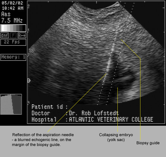

Image size:571 x 542px

In many cases, co-twins are not seen and pregnancy progresses. This usually leads to one of several outcomes. Some are shown here.

In the image below, a vesicle containing a degenerating structure believed to be an embryo, was seen on the chorion of its 60 day old co-twin. Judging from the size of this vesicle the conceptus was probably about 40 to 45 days old at the time of its death. The age of the surviving co-twin was not recorded but was probably about 65 days. 


Image size: 1569 x 986px

It is possible for a dead co-twin to co-exist with a live fetus until term. Occasionally, the small, mummified fetuses will be found within the placenta of the normal foal. In other cases, the pregnancy may not go to term. This is exemplified by the case illustrated below. The mummified fetus had obviously been dead for some time yet the surviving co-twin appeared to have been normal until shortly before it was aborted. Therefore, the reason for death was not obvious in this case. Indeed, it is tempting to suggest that abortion may have been due to an unrelated problem such as EHV1 infection.


Image size:1218 x 870px Modified from original. Copyright Copyright: Dr Cyril Stephen. Charles Sturt University. NSW.  Au. cyrilstephen@gmail.com

In most cases, the cause for aborted twin pregnancies appears to be related to the presence of its a co-twin. The reason for abortion is still poorly understood but may involved immunological incompatibility, endometrial  sharing and other mechanisms.  

Inset images A, B and C in the amalgam below show abortions that were probably related to the fact that they were twin pregnancies. These cases occurred many years ago, long before the use of ultrasound in stud practice and before the groundbreaking work of Dr O.J.Ginther, a pioneer in our understanding of twinning. Nowadays, with optimal stud management, similar images should be rare. 

In the last image (D) one can see the result of a twin pregnancy that progressed to term. Occasionally normal twins are born but most twins require intensive and expensive neonatal treatment. Even so, many of these foals die or are euthanized after prolonged treatment.


Image size: 1569 x 1989px

Interestingly, it is more common for the smaller neonate to survive than its larger co-twin. In that regard, it has been suggested that the smaller co-twin is under more stress than its co-twin, accelerating fetal maturation.  

It should also be noted that twin pregnancies are also potential causes of dystocia during abortion or during birth at term. In inset C, a "wryneck" fetal malformation in the larger foal complicated that delivery more than otherwise. 

A cautionary note:
Often a mare is presented with premature lactation; a strong indication that abortion is imminent. If the mare has not been under one's care from the time of conception, it is just as well to warn the owner that twins may be causing the abortion. Naturally, an effort should be made to exclude placentitis. Also, the owner's expectation as to the efficiency of EHV1 vaccination should be tempered with reality. However, the possibility of twins should not be discarded even if twin heart beats cannot be discerned on transabdominal ultrasonography on repeated examinations. 


Tuesday, November 13, 2018

Unusual placentation in a twin pregnancy. 

Keywords: placenta, equine, mare, inverted, twin, complication

Companion to an entry on twinning in mares in LORI.

There is still some uncertainty as to how and why twin equine pregnancies result in abortion. Sharing the available endometrial area is certainly a significant factor. Although typical inflammatory reactions at placental interfaces do not characterize twin abortions, immunological rejection of one co-twin by another may also be an important factor. Figure 1 shows one of numerous permutations of twin placentas apposed to one another within the uterus; placental sharing of the endometrial surface may be almost equal. In others, sharing is dramatically unequal. Figure is a schematic representation of such a case. Indeed, the situation in this pregnancy.

In Figure 1 red arrows indicate an area of apposition between the two chorionic surfaces. In these placentas there was little if any, macroscopic reaction between the two conceptuses despite the intimacy of placental apposition. Approximately 40% of the chorionic surface from the smaller co-twin was invaginated into the placenta of the larger twin.

Figure 1: Apposition of twin placentas and the path of delivery for both foals. Size available: 1626 x 2172px

The green arrows in Figure 1 show where the chorioallantois of the larger twin ruptured; in the region of the cervical star. The chorioallantois containing the smaller foal (yellow arrows) appeared within the allantoic cavity of the larger foal, invaginated within a pocket of the larger foal's placenta. The smaller foal was then delivered through a rupture of two layers of chorioallantois; that of the larger foal and that of its own placenta. The lower part of Figure 1 shows the path (grey arrow) that had to be taken by the smaller foal to be born; through the placenta of the larger foal.

Figure 2. A schematic representation of the placentation in this case. In some cases, the smaller co-twin may die and become a mummified attachment to the placenta of a normal foal. Image size available: 2553 x 1886px

Often, the placentas of twin foals will lie side by side within the uterus and each foal will be born in a conventional fashion, through the rupture of independent chorioallantoic membranes adjacent to the cervix.

Monday, December 30, 2013

Routine monitoring of early pregnancy

Keywords: equine, mare, pregnancy diagnosis, monitoring

This sequence of images illustrates the author’s philosophy in monitoring early pregnancies.  Mare owners are encouraged to submit their mares for all of these steps in diagnosis. In this manner, twins can be managed properly, early embryonal death can be detected and the normal development of the conceptus can be monitored. Note the centimeter scale in all images.

Image A shows a conceptus presumed to be about 14 days old.


Image size 461 x 313px (full size).

In image A again, the echogenic line (a line of strong spectral reflections) that characterizes the uterine body (arrow) shows that the embryo is just cranial to the cervix.

The precise age of an embryo is unknown in common stud practice because mares are examined and usually inseminated at two day intervals (Mon-Wed-Fri) or three days intervals (Fri-Mon). This means that the precise time of ovulation is usually unknown.
 
The embryonic vesicle migrates throughout the uterus until the embryo is about 16 days old then becomes "fixed". If there are twin embryos, they often become "fixed" together, complicating twin reduction; usually making it impossible. Therefore it is important to err on the side of shorter, rather than longer gestation when scheduling pregnancy diagnoses. 

Because the time of conception is usually unknown in common stud practice, pregnancy diagnosis is scheduled 14 to 16 days after an un-ovulated follicle was last seen, not after the first record of ovulation. In the latter case, twin embryos could be two or three days older than 14-16 days and already "fixed".


Image B shows a conceptus that is approximately 16 days old.


Image size 461 x 313px (full size).

It has already become "fixed" in the uterine horn. The thick homogeneous "doughnut" that surrounds it is the tonic myometrium that characterizes early pregnancy in this species. The non-echogenic (black) center of the vesicle is almost entirely yolk sac. The embryo itself is not yet visible.

Image C shows a pregnancy is approximately 19 days of age.


Image size 461 x 313px (full size).

The sodium pump in the wall of the trophoblast is less active than earlier in gestation therefore the osmotic pressure within the trophoblast is lower and the trophoblast loses some of its turgidity at this time. This is normal and should not lead one to assume that embryonal death is about to occur.  A small echogenic area on the lower right extremity of this vesicle  is the embryo itself (arrow).

In image D, the embryo (green arrow) has lifted away from the ventral portion of the trophoblast about 23 to 24 days of age.


Image size 461 x 313px (full size).

The characteristic thickening of the uterine wall opposite the embryo (yellow arrow) is typical of this stage of pregnancy.

In images E and F, a pregnancy about 27 days old, the embryo itself is clearly visible and the embryonic membranes can be distinguished as well.


Image size 461 x 313px (full size).

These structures are delineated in image F.


Image size 461 x 313px (full size).

The heartbeat of the embryo clearly visible at this time, usually in the range of 120 to 160 bpm.

Tuesday, May 12, 2015

Complications of a twin pregnancy: a video


Keywords: video, Thoroughbred, twins, complications, physiology, anatomy, placentation


To play the video, click on this link.

An excellent teaching case, incorporating many examples of physiology, management, pathology, anatomy, obstetrics and neonatology.

This video exemplifies the need to diagnose twins early in gestation. It covers two methods of reducing twins to singlets, shows how twins can be diagnosed or missed in late in gestation and demonstrates the need for inducing foaling in some cases. It also illustrates how to decide when induction should occur and how to induce foaling. A case of premature placental separation is illustrated, preceding the delivery of twin foals. There is also a bizarre variation of placental development. The case ends tragically with euthanasia of both foals as early neonates.

Wednesday, April 16, 2014

Genital tubercles, genitalia and gonads in fetal sexing

Keywords: fetus, sexing, ultrasound, male, equine

There are many reasons to determine fetal gender in horses. These include a multitude of personal decisions by owners such as preferences for stud Thoroughbred colts, the wish to retain fillies for breeding, insurance concerns, a need for female polo ponies, male draft horses and so on.

Detection and examination of the genital tubercles

First, a note on accurate age determination of an equine fetus. In general stud practice, determination of the exact duration of pregnancy is not possible. Mares are commonly examined at two day intervals or over weekend at three day intervals. When a CH is first detected, it is impossible determine its age. Therefore ovulation could have occurred shortly before one's examination or close to two or three days earlier. If one assumes a two day interval between examinations and pregnancy is timed from when the follicle was last seen, a "55 day pregnancy" could be 53 to 55 days old. If the duration of pregnancy is timed from when a CH is first seen, the pregnancy could be 55 to 57 days old. This seemingly trivial point takes on critical importance when a mare is found to be pregnant with twins at "15 days" of gestation!

It is only when frozen semen is used, that the approximate time of ovulation (and the age of the fetus) is known. Even when embryo transfer is used, the exact age of the fetus is not known. Therefore all the fetal ages in this discussion must be regarded as approximate.

Early sex determination is usually done by transrectal ultrasonography, between 59 and 68 days of gestation by detecting the genital tubercle. The genital tubercle is the precursor of the penis in the male and the clitoris in the female. Around day 55 gestation it appears as a hyperechoic equal sign (=) located between the fetal hindlimbs, at an approximately equal distance between the tail and the umbilicus. As the fetus grows the relative position of the tubercle changes, becoming closer to the tail in the female and the umbilicus in the male.

The image below shows a 64 day male fetus still in its amnion. The arrow indicates the position of the genital tubercle (arrow) used for sex determination. It is highly echogenic and usually easy to locate, just caudal to the umbilical cord. In females, the genital tubercle is located under the tail i.e. in the position of the vulva in a neonate.

In this case, the remnants of the yolk (YS) can be seen, consistent with this stage of gestation.


Image size: 1000 x 717px.

The image below shows the genital tubercles (green rings) of twin fetuses aborted at approximately 60 days of gestation. The upper fetus is male, the lower one female.


Image size: 1000 x 744px. 

Detection and examination of the external genitalia, mammary glands and gonads.


A wide diagnostic window for gender determination exists between 100 and 260 days gestation. During this time, the external genitalia, mammary glands and fetal gonads themselves are examined. and multiple parameters to validate diagnosis (fetal primary sex organs), but may require a combination of trans-rectal and trans-abdominal ultrasound scanning.

The external genitalia of the fetus may be clearly identified on ultrasound as early as 100 days gestation. In the male fetus, a fully comprehensive gender diagnosis includes the identification of the penis, prepuce scrotum/testicular compartments urethra and gonads. In the female fetus, the vulva and clitoris, mammary gland, nipples and again, the gonads themselves.

In the 150 day old male fetus below, the scrotum and penis are well formed but the testicles have not yet descended into the scrotum. Each enlargement seen within the scrotum in this fetus is due to the gubernaculum, not the testicle.  This explains why the scrotal contents are relatively anechogenic  during fetal sexing (see the image below this one).


Image size: 1000 x 757px. 5 x 3.285 inches 200dpi.

In this transabdominal ultrasonograph, captured at seven  months of gestation, the anechogenic contents of the scrotum and the penis are clearly visible. Note the relatively anechogenic scrotal contents.  The penis itself is not visible here because it runs on a plane that is dorsal to the urethra and scrotum.


Image size: 820 x 614px  Author and copyright. Dr Stephanie Bucca: Doha, Qatar. stefbucca@gmail.com

At four to seven months in gestation, the vulva lips, mammary gland and teats can be used to identify a female fetus.


Image size: 2050 x 1612px  Author and copyright. Dr Stephanie Bucca : Doha, Qatar. stefbucca@gmail.com

In image A, the base of the fetal tail is visible with faint cross sections of some of the coccygeal vertebrae seen to the left of the tail base. Between the buttocks of the fetus one can see its anus, vulva lips and clitoris.

In image B, at just under five months of gestation, this fetus is in posterior presentation showing one half of the mammary gland and its nipple. One  hind limb is extended, with the femur shown in cross section. Vernix is clearly visible in the amnion but not the allantois.

In image C, the fetus is in transverse dorsal presentation. Between its buttocks, the anus and vulva lips of the fetus are clearly visible. Essentially, this is the same view as in image A, but closer to the fetus

After about eight months or nine months of gestation, transabdominal ultrasonography for sexing is less predictable than earlier. Although the fetus becomes fixed in cranial longitudinal presentation, its hind quarters are encased in the pregnant horn and may be raised towards the ovaries. (The hind legs are often palpable per rectum in late gestation). In fact, at this time, fetal cardiac examination can be more fruitful than attempts to examine their genitals. Even with low frequency ultrasound, penetration may not be sufficient to examine genital areas of diagnostic interest.

Although male and female fetal gonads have a similar macroscopic appearance, they differ remarkably on ultrasonography, especially when examined by doppler flow ultrasonography.

On the right side of this image, the tunica albuginea and the clear demarcation between cortex and medulla is obvious. The medulla is peripheral to the cortex in equids (a quirk of embryology) and because of its blood supply, it is less echogenic than the cortex. The donut-like appearance of the fetal ovary is very different to the relatively homogeneous appearance of the fetal testicle (see the following image). This forms the basis for accurate sexing of equine fetuses. Approximate time windows for these examinations are between 4 and 5 months transrectally and 7 and 8 months transabdominally.


Image size: 2050 x 1612px  Author and copyright. Dr Stephanie Bucca: Doha, Qatar. stefbucca@gmail.co

When doppler flow ultrasonography is used, the accuracy of gender determination is close to 100% accurate. This because the medulla of the ovary is well vascularized, making it an ideal target for doppler flow examination. The value of doppler flow ultrasonography is easily appreciated in the image above (the scale is the same for both sub-images).

In male fetuses examined by doppler flow ultrasonography, the donut-like appearance is absent and doppler flow variation is largely restricted to the centrally located blood vessels. That appearance is shown here:


Image size: 820 x 614px  Author and copyright. Dr Stephanie Bucca: Doha, Qatar. stefbucca@gmail.co.

Some find doppler flow imaging less useful than others, deferring to the appearance of male and female gonads on B mode ultrasonography alone. This is because movement of the fetus itself can cause considerable echogenic noise during doppler flow ultrasonography.

The author wishes to acknowledge the assistance of  Drs Stephanie Bucca, Juan Samper and Carlos Pinto in creating this LORI entry.


Video (click on image and decrease page size to view at optimal resolution):


 Sexing video


Video (PENDING) size 614 x 480px. Assembled and edited by Dr Rob Lofstedt (lofstedt@upei.ca). Copyright; Dr Carlos Pinto (tubercle files) Carlos.Pinto@cvm.osu.edu and Dr Juan Samper (gonad files) jsamper@telus.net. 

References:

1. Bucca S. 2005. Equine fetal gender determination from mid- to advanced-gestation by ultrasound. Theriogenology 64:568–571

2. Renaudin, C.D., Gillis C.L. and Tarantal, A.F.  1997 Transabdominal combined with transrectal ultrasonographic determination of equine fetal gender during Midgestation. AAEP proceedings. 43: 252-255.

3. Resende H.L. et al. 2013. Determination of equine fetal sex by Doppler ultrasonography of the gonads.Equine Vet J. 2013 Nov 15. doi: 10.1111/evj.12213. (Epub)

Tuesday, February 19, 2019

The oviduct (uterine tube) revisited

Keywords: equine, mare, oviduct, uterine tube

The Nomina Anatomica Veterinaria refers to this structure as the uterine tube (to distinguish it from the oviduct of birds). However, that term is seldom used in either practice or publication. In both humans and animals, it is instead referred to as the oviduct. The oviduct in any species is amazing but even more so in mares. This entry substantiates that impression.

As shown in figure 1, the oviduct runs within the ovarian bursa, almost parallel to its margin but a full centimeter or more away.

Figure 1. The left ovary of a two year old mare, suspended under water. The ovulation fossa is invisible, pointing ventrally in the image. 2903 x 2054 px


Figure 1 is labeled above. Divisions of the oviduct (infundibulum, ampulla etc) shown here are those adopted from, and and described in: Aguilar, J.J. et al. 2012. Histological characteristics of the equine oviductal mucosa at different reproductive stages. J.Equine.Vet. Sci. 32:99-105. Note: The ovary in situ hangs from the mesovarian ligament so that the ovulation fossa faces ventrally. In this image, the ovary and bursa have been rotated as shown by the arrow in the small inset. The ovulation fossa is not yet visible despite this rotation. Image size: 1600 x 1041 px

The ovarian bursa can be likened to a lateral, low-drooping eyelid over an eye (the ovary). The long dimension of the ovoid-shaped ovary lies on a cranial-caudal axis. The cranial pole of this axis is slightly higher than the caudal pole. The infundibulum lies at the cranial pole of the ovary. As shown in figure 1, it is not attached to the ovary. In fact, it lies a remarkable distance from the ovulation fossa.

This anatomy never ceases to amaze the author. In essence, the infundibulum acts like a catcher's mitt in a baseball game, covering an ambitious area some distance from the pitcher's mound i.e. the ovulation fossa in this analogy. The baseball is of course, the oocyte. The infundibulum is well supplied with smooth muscle and engorged blood vessels during estrus, expanding the catcher's mitt. Yet the precise mechanism and magic behind the dependability of the catcher remains unknown and unseen.

For those not familiar with baseball i.e. (insert nationality here), the author suggests consulting the book "Complete idiots guide to baseball and oocytes".

It has been suggested that the fimbriae of the infundibulum sweep the surface of the ovary at the time of ovulation, picking up the oocyte in the process, moving it into the complex folds of the infundibulum. Although the frequency of loss of oocytes into the peritoneal cavity is unknown in mares, it probably does occur; it has certainly been documented in humans. Certainly, losses of oocytes into the peritoneal cavity is described in poultry, especially broiler hens. Interestingly, laying hens, selected for egg production are less prone to peritoneal loss of oocytes. In rodents and canids, oocyte-catching expertise by the infundibulim is less important than other species. This is because ovarian bursae in those animals surround their ovaries completely and are continuous with the infundibula themselves. This makes it impossible for their oocytes to escape into the peritoneal cavity.

Figure 2: An oocyte, 150µ in diameter is shown at the end of the yellow arrow. This simulates the appearance of a real oocyte shortly after ovulation. Note its size relative to the infundibulum and ostium. Also note the cloud-like mass around the oocyte. This is a simulation of the large cumulus oophorus that accompanies the oocyte into the infundibulum. The cumulus is lost within 6 to 12 hours after the oocyte enters the oviduct (personal communication; Dr Katrin Hinricks).  Image size: 1630 x 1236 px

When stretched out, the oviduct in a mare is a little longer than the human hand i.e. about 20 to 30 centimeters. It is a continuum with no distinct delineation. To facilitate functional descriptions however, it is divided into three main sections i.e. the infundibulum (L.< funnel), ampulla (L.< flask) and finally the isthmus (L.<neck of land between two seas) narrowing as it enters the uterus at a papilla that forms the uterotubal junction.

Figure 3: This figure includes a large section of the infundibulum and a smaller inset image of the isthmus. They are both at the same scale of magnification. Again, the author has modeled an oocyte in the infundibulum and in addition, an embryo in the isthmus. Both are visible beside black bars 150µ ling at the end of the yellow arrows. This was done to compare the size those structures with the histology of parts of the oviduct. The diameter of an equine oocyte is approximately 150µ; slightly larger than that a bovine oocyte (120µ). By day 6, the equine embryo is approximately 200µ in diameter. The white scale within each image is 500µ and the small bar adjacent to the oocyte and embryo is 150µ long. Image size: 4242 x 3090 px

The oviduct is of course, a conduit to transport oocytes, then embryos from the ovary to the uterus. But it is also an organ that performs the seemingly impossible task of (often simultaneously) transporting oocytes towards the uterus while promoting the ascent of spermatozoa from the uterus into the oviduct. Like other tubular organs throughout the body it has inner circular and outer longitudinal layers of smooth muscle that promote peristaltic movement. It also has within its mucosal lining, ciliated cells that also play a role in gamete transport. Presumably, peristaltic movements play a major role in transporting oocytes towards the uterus while cilia perform a major role with regard to the ascent of spermatozoa. However, the exact integration of these two propelling mechanisms has yet to be described.

It is known that spermatozoa ascend into the oviduct and bind to its epithelium, usually laying in wait for the oocyte to arrive after ovulation. It is possible for fertilization to occur when spermatozoa arrive in the oviduct up to 18 hours after ovulation (with post ovulation insemination) but it is far more common for spermatozoa to spend a day or two or even up to 7 days in the oviduct before ovulation. During that time, Ca++ fluxes within the oviduct suppress capacitation while gaseous exchange and nutrition keep spermatozoa viable. True, it is more likely that an oocyte will be fertilized with close synchrony between insemination and ovulation but the ability of the oviduct to keep spermatozoa viable for long periods of time is still remarkable; far superior than any device contrived by humans.

As amazing as sperm preservation maybe in mares, it is overshadowed by the achievement of oviducts in other species. In some fruit bats in hibernation for example, spermatozoa can survive for weeks even months within the oviduct! 

Capacitation and the release of spermatozoa from their binding sites on the oviduct epithelium is orchestrated by changes in the steroid milieu, especially increased progesterone production shortly before ovulation. The effect of the oviduct environment on spermatozoa is of critical importance in mares. Specifically (and peculiar to mares again) it is only in the oviduct that fertilization can occur. Therefore, unless spermatozoa are injected directly  into oocytes (ICSI), in-vitro fertilization in horses is seldom successful.


Figure 2: A 16 gauge blunted needle is introduced into the ostium of the oviduct from a two-year-old mare. Blue dye is then introduced to outline the convoluted shape of the oviduct. Evidence of the dye in the uterus is shown as it permeates through to the serosa at the site marked B. This image defines the oviduct clearly. However the anatomical divisions (especially the ampullary-ithmic junction) so glibly discussed in literature, are far from obvious. Image size: 3456 x 2557 px

The uterotubal junction too, is a remarkable structure in mares. Spermatozoa deposited in the uterus are swept up to the uterotubal junction and are found in the oviduct within a few minutes of insemination. Yet, it is virtually impossible in normal mares, to force either fluid or air from the uterine lumen into the oviduct. This is because the oviduct of the mare is unique amoung domestic species with respect to its uterotubal junction. In mares, the distal oviduct has a well developed muscularis which acts as a sphincter, making mechanical entry from the uterus difficult. One can introduce fine tubes into the oviduct from the uterus but otherwise the uterotubal junction in mares acts as a one-way valve preventing fluid ascent from the uterus. To some degree, this may explain the relative lack of oviduct pathology in mares compared to cattle.

Alter about six and a half days in the oviduct, incubating mainly at the ampullary-ithmic junction, embryos enter the uterus. In mares of course, single embryos are far more common than twin embryos. At that time, late morulas or early blastocysts can be collected by flushing the uterus. Before that time it is impossible to retrieve a fertilized embryo by flushing the uterus alone.

Although unfertilized oocytes are occasionally found in the uterus, this is unusual. In general, if an oocyte is not fertilized in mares, it will not reach uterus. This phenomenon is unique among equids. Therefore it is generally not important to determine if oocytes have been fertilized when they are collected for embryo transfer. It is now universally recognized that production of prostaglandin E2 by embryos (not oocytes) causes relaxation of oviduct smooth muscle. This allows transport of the embryo into the uterus. When mares are examined postmortem, it is not unusual to find flattened, degenerate oocytes from previous cycles, caught within the oviduct.

Pathology? 
Fibrinous masses that can be several mm in size, are often found in the oviducts of mares. Again, this is a phenomenon peculiar to equids. It has been suggested that they are pathological and may block the passage of  oocytes and embryos in the oviduct. However, these masses are found in 75% to 85% of mares (as reviewed by Tsutsumi, Y. 1979) therefore they are unlikely to be pathological. The origin of the masses is unknown but they may arise from fibrin discharged from follicles after ovulation, during the formation of corpora hemorrhagica. In that regard, it is also very common to see fibrin tags in and around the ovary in apparently normal mares. In fact, careful inspection of the images in this entry will reveal such tags. The author had the dubious privilege of spending many hours at an equine slaughter plant and saw such tags many times, often in young mares. The same can be said for para-ovarian (wolffian) cysts, sometimes reported as abnormal too. The vast majority of otherwise normal mares have these cysts. 

Selected references:

Allen, W.E. et al. 1979. Evaluation of uterine tube function in pony mares. Vet. Record 105: 364-366

Arnold, C.E. and Love, C.C. 2013. Laparoscopic evaluation of oviductal patency in the standing mare. Theriogenology 79: 905-910

Bennett, S. 2002. Surgical evaluation of oviduct disease and patency in the mare. Proc. AAEP 48:347-349

Betteridge, K.J. 2000. Comparative aspects of equine embryonic development. Anim. Reprod. Sci. 60: 691-702

Brinsko, S,P. 1991. The effect of uterine lavage performed four hours post insemination on pregnancy rate in mares. Theriogenology 35: 1111-1119

Dobrinski, I. et al. 1997. Membrane contact with oviductal epithelium modulates the intracellular calcium concentration of equine spermatozoa in vitro. Biol. Reprod. 56: 861-869

Freeman, D.A. 1991. Time of embryo transport through the mare oviduct. Theriogenology 36: 823-830

Ghazal, S et al. Glob. libr. women's med., (ISSN: 1756-2228) 2014; DOI 10.3843/GLOWM.10317

Hinrichs, K. 2010. In vitro production of equine embryos: State of the art. Reprod. Domestic Anim 45: 3-8

Hunter, R.H.F. 1999 Ovarian follicular fluid, progesterone and Ca2+ ion influences on sperm release from the Fallopian tube reservoir. Gamete biology. 54: 283-291

Hunter, R.H.F. 2008. Sperm release from oviduct epithelial binding is controlled hormonally by peri‐ovulatory graafian follicles. Molecular Reprod. Devel. Incorporating Gamete Research 75: 167-174

Inoue, Y. 2013 Hysteroscopic hydrotubation of the equine oviduct. Equine Vet J. 45:761-765

Kenney, R.M. 1993. A review of the pathology of the equine oviduct. R. M. Kenney. Equine Vet. J. 25 (S15): 42-46

Leemans, B.M. 2015. Why doesn’t conventional IVF work in the horse? The equine oviduct as a microenvironment for capacitation/fertilization. Reproduction 152: R233-R245

Navara, K. J. 2015. Higher rates of internal ovulations occur in broiler breeder hens treated with testosterone. Poult Sci. 94:1346-1352

Rigby, S. et al. 2000. Oviductal sperm numbers following proximal uterine horn or uterine body insemination. Proc. AAEP. 46:332-334

Saltiel, A. et al. 1986. Pathologic findings in the oviducts of mares.  Am. J. Vet Res. 47: 594-597

Sieme, H. et al. 2003. The effects of different insemination regimes on fertility in mares. Theriogenology 60: 1153-1164

Smits, K et al. 2016 The equine embryo influences immune-related gene expression in the oviduct. Biol. Reprod. 36: 1-8

Tsutsumi, Y. 1979. Evidence of the origin of the gelatinous masses in the oviducts of mares.
J. Reprod. Fert. 57: 287-290

Weber, J.A. 1995. Relaxatory effect of prostaglandin E2 on circular smooth muscle isolated from the equine oviductal isthmus. Biol. Reprod. Monograph. series1: 125-130