EQUINE IMAGES

How to use these images


IMPORTANT: To see an enlarged image, click on any image you see in LORI. Then, RIGHT click on the enlarged image to save it at its full size.


Showing posts with label mare. Show all posts

Tuesday, November 13, 2018

Unusual placentation in a twin pregnancy. 

Keywords: placenta, equine, mare, inverted, twin, complication

Companion to an entry on twinning in mares in LORI.

There is still some uncertainty as to how and why twin equine pregnancies result in abortion. Sharing the available endometrial area is certainly a significant factor. Although typical inflammatory reactions at placental interfaces do not characterize twin abortions, immunological rejection of one co-twin by another may also be an important factor. Figure 1 shows one of numerous permutations of twin placentas apposed to one another within the uterus; placental sharing of the endometrial surface may be almost equal. In others, sharing is dramatically unequal. Figure is a schematic representation of such a case. Indeed, the situation in this pregnancy.

In Figure 1 red arrows indicate an area of apposition between the two chorionic surfaces. In these placentas there was little if any, macroscopic reaction between the two conceptuses despite the intimacy of placental apposition. Approximately 40% of the chorionic surface from the smaller co-twin was invaginated into the placenta of the larger twin.

Figure 1: Apposition of twin placentas and the path of delivery for both foals. Size available: 1626 x 2172px

The green arrows in Figure 1 show where the chorioallantois of the larger twin ruptured; in the region of the cervical star. The chorioallantois containing the smaller foal (yellow arrows) appeared within the allantoic cavity of the larger foal, invaginated within a pocket of the larger foal's placenta. The smaller foal was then delivered through a rupture of two layers of chorioallantois; that of the larger foal and that of its own placenta. The lower part of Figure 1 shows the path (grey arrow) that had to be taken by the smaller foal to be born; through the placenta of the larger foal.

Figure 2. A schematic representation of the placentation in this case. In some cases, the smaller co-twin may die and become a mummified attachment to the placenta of a normal foal. Image size available: 2553 x 1886px

Often, the placentas of twin foals will lie side by side within the uterus and each foal will be born in a conventional fashion, through the rupture of independent chorioallantoic membranes adjacent to the cervix.

Monday, March 2, 2015

Twins and infertility

Keywords: equine, infertility, twins, ovulation, abortion.

Note: the term conceptus is used here by the author, in preference to embryo. In the author's mind, conceptus refers to the embryo together with its membranes while embryo refers to the future fetus. Also, because of the importance of twinning in equine reproduction, the author has included more text than usual in this entry.

Twin preovulatory follicles seen on ultrasound.


Image size: 900 x 694px

Conventional wisdom once dictated that mares should not be bred when twin preovulatory follicles were present. This was based on the fact that twins are a common cause of abortion in mares. With the increase in knowledge that came with common use of ultrasonography, this philosophy has changed. Today, it would be considered poor management to miss this breeding opportunity. Indeed, if mares where not to be bred whenever twin follicles were present, perhaps 15 to 20% of all breeding opportunities would be lost. This is especially the case in breeds such Thoroughbreds and Standardbreds where twin ovulations are common. It is also significant in any breed with the approach of the summer solstice because day length and ovulation rate are positively correlated.

Current philosophy dictates that mares should always be bred when twin follicles are present. Indeed, even when twin pregnancies are not discovered, the overall singlet pregnancy rate is higher when this approach is used.


Notes on the timing of pregnancy diagnosis:

When two conceptions occur during a single estrous period, one conceptus will invariably be older and larger than the other conceptus when pregnancy diagnosis is performed. This is important to bear in mind. Although not common, twin ovulations can be separated by several days, making it possible to miss a younger conceptus when routine pregnancy diagnosis is performed at 14 to 15 days after ovulation. 

Newer, high resolution ultrasound units make it possible to see pregnancies at 12 days or even earlier. If there a second, younger conceptus in the uterus at this time, it may not be seen. However, if pregnancy diagnosis is delayed to 14 days, it is very likely that both conceptuses will be seen. After 16 days, embryo movement within the uterus will have ceased and the opportunity to crush one embryo without harming the other will have diminished significantly. Therefore pregnancy diagnosis should be scheduled at 14 to 16 days after the time that the first of two intact (preovulatory) follicles was last seen. This is explained below:

On many stud farms, visits are made on Mondays, Wednesdays and Fridays. If the time of ovulation is taken as the time a corpus luteum is first seen on ultrasonography, it can amount to a critical error in timing of pregnancy diagnosis. This is because the actual time of ovulation could have been two or even three days earlier, immediately after one last saw an intact follicle.

Finally, two large follicles may be present yet, with the vagaries of ultrasonography and work pressure, it is not rare for one to be under the impression that there is only a single pre-ovulatory follicle. Therefore one should always assume that twins may be present unless proven otherwise!


Taken 14 to 15 days after the last record of an intact follicle, the images below show how quickly embryos can migrate within the uterus before the time of fixation. This is used to one's advantage when crushing one of the embryos (usually the smaller of the two).


Image size: 1800 x 1369px

Movement through the uterus is a product of myometrial contraction i.e. even inanimate objects will move quickly within a mare's uterus. This is easily demonstrated by inserting small, sterilized fluid- filled "balloon twins" into the uterus via the cervix.


Image size: 1308 x 905px

Movement of embryos is thought to be important in the recognition of pregnancy but stops abruptly at about 16 days after ovulation.

In practical terms, it is very important for the veterinarian to examine every part of the uterine horns and body before excluding the possibility of twins.  Sometimes one of the twins may be present in a uterine horn while its co-twin is found in the uterine body, just cranial to the cervix.  It is quite easy to miss a co-twin in the uterine body if it is slightly off-center. Care must be taken to ensure that the specular reflection of the endometrial surface is visible as a thin echogenic line along the entire length of the uterine body from cervix to bifurcation. An illustration of this echogenic line can be seen in first image of this LORI entry

As mentioned, it is common to crush one co-twin to prevent abortion of both fetuses later in gestation. After tranquilization and rectal relaxation (n-butylscopolamine) as needed, the author locates the co-twin to be crushed and withdraws the transducer. The conceptus is then crushed immediately (before it is moved by the myometrium) as shown below. If the crush has been successful, little or no fluid will remain visible. Occasionally, several attempts at crushing may be required.


Image size: 800 x 552px

If there is concern that attempts at crushing have released enough prostaglandin to cause luteolysis, altrenogest (Regumate™) should be administered until the accessory corpora lutea have formed and the serum progesterone concentration has risen to well over 2ng/ml. Fortunately, altrenogest does not cross-react with most progesterone assays, so endogenous progesterone concentrations can be monitored until it is safe to wean the mare off altrenogest treatment.  

If one fails to crush a co-twin or if pregnancy diagnosis has been delayed beyond 16 days, other methods of twin reduction must be adopted. If a co-twin is crushed after this time, it is likely that both conceptuses will die; the reasons for that being beyond the scope of this discussion. Twin reduction using trans-vaginal ultrasound guided aspiration can be successful in these cases and should be attempted in favor of crushing if the critical day 16 has passed.

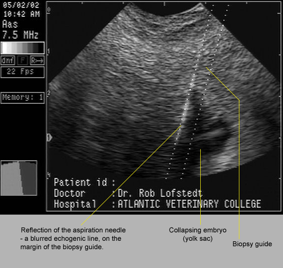
In the image below, 17 day old twins had fixed close to one another within the uterus. One was punctured and drained by ultrasound-guided aspiration through the fornix of the cranial vagina. If one has access to the correct equipment, this is a straightforward procedure.


Image size:571 x 542px

In many cases, co-twins are not seen and pregnancy progresses. This usually leads to one of several outcomes. Some are shown here.

In the image below, a vesicle containing a degenerating structure believed to be an embryo, was seen on the chorion of its 60 day old co-twin. Judging from the size of this vesicle the conceptus was probably about 40 to 45 days old at the time of its death. The age of the surviving co-twin was not recorded but was probably about 65 days. 


Image size: 1569 x 986px

It is possible for a dead co-twin to co-exist with a live fetus until term. Occasionally, the small, mummified fetuses will be found within the placenta of the normal foal. In other cases, the pregnancy may not go to term. This is exemplified by the case illustrated below. The mummified fetus had obviously been dead for some time yet the surviving co-twin appeared to have been normal until shortly before it was aborted. Therefore, the reason for death was not obvious in this case. Indeed, it is tempting to suggest that abortion may have been due to an unrelated problem such as EHV1 infection.


Image size:1218 x 870px Modified from original. Copyright Copyright: Dr Cyril Stephen. Charles Sturt University. NSW.  Au. cyrilstephen@gmail.com

In most cases, the cause for aborted twin pregnancies appears to be related to the presence of its a co-twin. The reason for abortion is still poorly understood but may involved immunological incompatibility, endometrial  sharing and other mechanisms.  

Inset images A, B and C in the amalgam below show abortions that were probably related to the fact that they were twin pregnancies. These cases occurred many years ago, long before the use of ultrasound in stud practice and before the groundbreaking work of Dr O.J.Ginther, a pioneer in our understanding of twinning. Nowadays, with optimal stud management, similar images should be rare. 

In the last image (D) one can see the result of a twin pregnancy that progressed to term. Occasionally normal twins are born but most twins require intensive and expensive neonatal treatment. Even so, many of these foals die or are euthanized after prolonged treatment.


Image size: 1569 x 1989px

Interestingly, it is more common for the smaller neonate to survive than its larger co-twin. In that regard, it has been suggested that the smaller co-twin is under more stress than its co-twin, accelerating fetal maturation.  

It should also be noted that twin pregnancies are also potential causes of dystocia during abortion or during birth at term. In inset C, a "wryneck" fetal malformation in the larger foal complicated that delivery more than otherwise. 

A cautionary note:
Often a mare is presented with premature lactation; a strong indication that abortion is imminent. If the mare has not been under one's care from the time of conception, it is just as well to warn the owner that twins may be causing the abortion. Naturally, an effort should be made to exclude placentitis. Also, the owner's expectation as to the efficiency of EHV1 vaccination should be tempered with reality. However, the possibility of twins should not be discarded even if twin heart beats cannot be discerned on transabdominal ultrasonography on repeated examinations. 


Saturday, February 21, 2015

Mesenteric hemorrhage associated with foaling

Keywords: hemorrhage, foaling, equine, mare, intestine

A mature Standardbred mare that died suddenly immediately after foaling.  Her mucous membranes were extremely pale upon presentation and postmortem examination revealed that she had suffered from an extensive intra-abdominal hemorrhage. Contrary to expectation, the hemorrhage did not arise from a rupture of the uterine artery (prev. middle uterine artery). Instead, hemorrhage originated from one of the major vessels in the spleno-colic ligament. Bleeding extended into the mesentry and peritoneal cavity.


Image size: 1500 x 1227px

Despite an adequate worming program on this stud farm, the presiding pathologist suggested the mare could have experienced verminous arteritis and by extension, arterial rupture because of hypertension during foaling. However, some doubt  has been leveled at that suggestion, based on the fact that verminous arteritis due to S. vulgaris is still well controlled by current anthelmintics and cases of arteritis are no longer common in this geographical area (N.E. Canada). Instead, it has been suggested that the trauma seen here may have been sustained by the foal during parturition. The accompanying review substantiates that idea.

Reference: 
Dolente, B.A. et al. 2005. Mares admitted to a referralhospital for postpartum emergencies: 163 cases (1992-2002). J.Vet.Emergency and Critical Care 15: 193-200

Friday, February 20, 2015

Third-degree perineal lacerations

Keywords: laceration, degree, mare, foaling, injury

First and second degree perineal lacerations are seldom of importance, the first being laceration of the vaginal mucosa and the second, laceration of the vaginal mucosa and the tissue between the vagina and rectum.

Third degree lacerations involve the vaginal mucosa, rectal mucosa, and the interposing tissue.  They may take the form of fistulas or more commonly, complete destruction of the perineal body. As mentioned in an entry on evisceration after foaling these accidents appear to be more common in maiden mares than pluriparous mares.  These old but valuable images illustrate the problem well.

In the top right inset of the image below, a third degree laceration is being examined several days after foaling. A large hematoma is present in the left wall of the vestibule and there is considerable fecal contamination over the lacerated tissue between the vagina and rectum.  These injuries are almost never fatal and hemorrhage is seldom severe because of the blunt nature of this trauma.

The author estimates that the main image was obtained perhaps ten days later, when the hematoma had resolved and secondary intention healing of the torn tissue was well underway.


Image size: 840 x 767px  Modified. Original copyright Dept. Theriogenology, ISU. Ames, Iowa.

Although the value of antibiotic treatment is debatable in these cases, immediately tetanus prophylaxis is suggested. Although there is some disagreement on when surgery should occur, the author suggests that one should resist the temptation to repair these injuries surgically until second intention healing is complete. If that is done, the wound margins and viable tissue can be clearly delineated for surgery.

In practice, the foal is also weaned before surgical repair is attempted because dietary restrictions imposed on the mare (to decrease fecal production) will retard the growth of the foal.

Owners may be concerned to wait for several month for repair because fecal contamination of the vagina continues during that period. Remarkably,this approach does not appear to have a detrimental effect on the future reproductive capacity of mares. This could be due to excellent uterine defense mechanisms in young mares,

Method of repair:


Image size: 743 x 824px  Modified. Original copyright Dept. Theriogenology, ISU. Ames, Iowa.

The author has used an Aanes two-stage technique modified by Dr. Tracy Clark at Iowa State University. The image at upper left shows how the perineum has been prepared for surgery. Pre-surgical starvation diminishes the amount of feces in the rectum (ringed in the lower left inset) at the time of surgery. After all feces have been removed by hand, fecal debris can be prevented from entering the surgical field by using a large tampon device made of rolled cotton, held together with half hitches of umbilical tape, complete with a tail of tape to withdraw it from the rectum after surgery.  Epidural anesthesia is used.

The inset image at lower right shows how the rectum, vagina and interposing tissue are repaired using a continuous, cranial-to-caudal suture pattern. No attempt is made to reconstruct the perineal body during first stage repair. Doing so will cause feces to exert pressure on the anus, breaking the suture line described above.

After the first stage has healed, the perineum is reconstructed.

Third degree lacerations of the perineal body may not involve the anus; after healing they take the form of  fistulas between the vagina and rectum. Sometimes these fistulas are large, allowing these animals to defecate through their vulva lips. Such a case is shown below.


Image size: 444 x 1050px  Modified. Original copyright Dept. Theriogenology, ISU. Ames, Iowa.

The lower image shows the area being prepared for surgery with a dramatic demonstration of the extent of the lesion to be repaired. The technique used is similar to the first stage described above.

Reference: 
Aanes, W.A. 1988. Surgical management of foaling injuries. Vet. Clin. North Am. Equine Pract. 4:417-438. 

Per rectal evisceration after foaling

Keywords: trauma, equine, mare, foaling, rectum

The first image shows a six year old mare presented shortly after her first foaling.  Despite video surveillance, foaling was rapid and consequently, unattended. The birth of a live foal was followed immediately by evisceration from the anus. As shown in inset A, a temporary purse string suture had been placed around the anus by the rDVM.


Image size: 990 x 747px

When the purse string was released, intestines burst out of the anus as shown in inset B. The placenta remained hanging from the vulva lips. The intestines had passed out of the peritoneal cavity through a tear in the rectum. Blood on the operator's glove after detection of the rectal tear:


Image size: 800 x 600px

The injury to the rectum was not caused by the foal because neither the uterus nor the vagina were damaged. This defies a simple explanation. It is possible that the rectum may have ruptured due to high abdominal pressure during foaling but the author is not aware of previous reports of such an accident. The mare was euthanized

More commonly evisceration occurs through vaginal tears.  The image below show how viscera can escape through the vagina. Note that the cervix and even the cranial vagina lie well within the peritoneal cavity, cranial to the reflection of the peritoneum in the recto-genital pouch. This anatomy makes it possible to perform ovariectomy via colpotomy in both mares and cows. However it becomes a potential liability in mares during foaling. This is because foaling is far  rapid than calving and a foal's forelimbs are more likely to traumatize the cranial vagina than is the case for calves. This is especially so if the foal is in foot-nape posture during second stage. If the foal’s hooves penetrate the vagina where it lies within the peritoneal cavity, intestines can enter the vagina through the site of penetration.

The image below clarifies this. LUH and RUH are the left and right uterine horns. PR is the peritoneal refection, edged by a red line, in the recto-genital pouch. The green lines show the cranial and caudal extents of the cervix. The red arrow points to a common site of vaginal penetration, the roof of the cranial vagina. The curved yellow arrow shows how intestines can move into the vagina. The rectum can be seen as a shaded body above the genital tract.


1232 x 919px

In the image below, a foal's legs penetrated the cranial vagina, entering the recto-genital fossa. This allowed the intestines (yellow arrow) including the large colon, to escape from the peritoneal cavity. Such cases are invariably fatal.


Image size: 1200 x 1008px Copyright Dept. Theriogenology, ISU. Ames, Iowa.

It is important to note that evisceration may occur some time after the birth of the foal, when the mare lies down and pressure is exerted on the abdomen. Evisceration through tears in the uterus is also possible, through the open cervix. Both vaginal and uterine tears can be present in mares that appear to be totally normal.  Although some owners object to potential interference with dam-offspring bonding immediately after foaling, the author always requests permission to examine the cranial vagina and uterus (as far cranial as possible) at that time.

The reader should be aware that it is far more common for a foal's hooves to tear through the caudal part of the vagina than the cranial part of the vagina. Tears often occur through the dorsal wall of the vestibule and there may even be total ablation of the perineal body. In those cases there is no danger of evisceration.

Note: Foaling accidents appear to be more common in maiden mares than pluriparous mares. This may be because the tract is smaller and less distensible in younger, than older animals. Also, maiden mares may panic and strain excessively during this novel experience. 

Saturday, January 10, 2015

Normal edema at term and ventral rupture

Keywords: ventral, edema, rupture, equine, mare, foaling, pain

The plaque of ventral edema shown below (arrows) is typical of late gestation in mares.


Image size: 1200 x 890 px

The affected area pits on pressure but the mare shows no discomfort at all. This makes this condition very different from impending rupture of the ventral abdominal structures.  That condition is shown below.

Impending rupture of ventral abdominal structures during late gestation.

A massive ventral plaque of edema (more obvious than the usual ventral edema of late gestation) and the painful gait of the mare are indicators of this condition. As shown in inset A, ventral edema may be so severe that the architecture of the mammary gland is largely obscured. Severe edema is also illustrated at lower left where the margin between the plaque of edema and the ventral abdomen is indicated by red arrows.


Image size: 1500 x 2102 px

Monitoring of milk electrolytes should be used to decide when to induce foaling. If the mare is off site, milk for electrolyte analysis can be collected by the owner. Assistance may be required during foaling. It has been suggested that minimal intervention is perhaps better that induction of foaling but that decision depends on prevailing conditions. In other words, if the mare is at a referral institution where 24 hour monitoring is possible, it may not be advisable to induce foaling. That is because induction of foaling is potentially hazardous, especially if it is done prematurely. In such cases, one would expect a higher incidence of failure of passive transfer and because the foal is not mature, a higher incidence of dystocia as well (movement into the correct position and posture is largely a function of fetal maturity).

After foaling, ventral edema usually subsides quickly enough to allow suckling (inset B). Otherwise, plasma transfusions may be given to prevent failure of passive transfer in the foal

If possible, the mare's abdomen should be supported until she can be induced to provide a viable foal. Supports can be made from wide elastic bandage i.e. Elastoplast TM, or a similar adhesive or partially adhesive bandage. Alternatively, a custom made support system can be used, such as that shown here.


Image size: 1200 x 1000px. Copyright Dr G. Colbern.  gtcolbern@gmail.com

Although the condition is often referred to as "rupture of the prepubic tendon" antemortem examination using ultrasound and posrtmorten examination in euthanized mares has shown that rupture may involve the prepubic tendon, the rectus abdominus muscle, the transverse abdominal muscles or oblique abdominal muscles.

Surgical reconstruction of the abdomen has been described but the long term success of those cases as broodmares is not known.  Soon after foaling, damage to abdominal is no longer visible so it may be tempting to re-breed these mares. This is not recommended because the condition is known to  reoccur in subsequent pregnancies.

References:

Blanchard, T.L. et al. 1987. Hydroallantois in two mares. Equine Vet J. 222-225

Ross, J et al. 2008. Body wall tears during late pregnancy in mares: 13 cases (1995–2006) J Am Vet Med Assoc. 232:257–261

Friday, December 5, 2014

Severe skeletal malformation a cause of dystocia

Keywords: dystocia, equine, mare, wryneck, wry-neck

A pluriparous mare presented for dystocia of several hours duration. The referring veterinarian reported the presence of "a severely deformed" foal that precluded normal delivery.


Image size: 1500 x 1949px

An epidural was given and clenbuterol were administered IV.

The foal was in anterior longitudinally presentation and dorso left-ilial position (arrow). There was an obvious wry-neck. Wry-neck is permanent ossification of the neck in a flexed posture; apparently unique to the horse among domestic animals.  The head was also deformed. Initially, one gained the impression that there was also bilateral shoulder flexion with both forelimbs retained.

Using a fetotome, the head was removed uneventfully but when an attempt was made to locate the forelimbs, they appear to be absent.  Because of the absence of the forelimbs, uterine tone and an inability to rotate the foal into a dorso-sacral position, the foal was delivered by cesarean section.  Her recovery was uneventful.

Radiology showed that both the left and right scapulae were present but only the right forelimb was present. Also, the right forelimb was vestigial, about the thickness of a thumb and it was also rotated on its longitudinal axis, so that the hoof faced backward. There was severe scoliosis and the head of the left femur did not articulate with the acetabulum.


Saturday, May 24, 2014

Cervical tears

Keywords: cervix, tear, equine, mare

Cervical tears often arise as a result of foaling, especially in maiden mares.  When the cervical integrity is sufficient to protect the uterine environment during pregnancy, cervical tears do not affect fertility. However, in many cases they do so; mares fail to conceive or conceive then experience early embryonic death.


Image size:1257 x 1320px

The mare with this cervical tear (top left) was presented after a failure to repair it surgically. This is not unusual because surgical repair of the cervix is difficult and healing is usually compromised because of its relatively poor blood supply.  The mare was infertile but her endometrial biopsy indicated that her uterus should be able to maintain pregnancy. Therefore a Shirodkar-like suture (a circumferential suture used for cervical closure in humans) was placed in this cervix and the mare was in inseminated at two successive estrous periods.  However ultrasonography failed to show pregnancy on both occasions at 14 days..

This illustration also attempts to emphasize that examination using either Polanski or tubular speculums can fail to demonstrate cervical tears.  This is especially the case during estrus when the cervix is relaxed. Even during diestrus, when cervical architecture is well defined, this author was unable to diagnose cervical tears on two occasions using speculum examination. Gloved hand examinations revealed the tears and their severity in both cases.

Thursday, April 24, 2014

Equine placental gross anatomy

Keywords: equine, mare, placenta, anatomy, yolk sac

The placenta of a normal equine term fetus. A clear plastic tube containing food dye has been inserted into the urachus. Note that the color of the dye is clearly visible through the wall of the umbilical cord. Although the urachus is lined with transitional epithelium (urothelium) that is continuous with the fetal bladder, it is little more than a space between the major vessels that occupy this portion of the cord. Therefore it cannot be isolated as a distinct vessel for surgical ligation when a neonate has a persistent urachus. Only if it is infected, inflamed and thickened will it attain substantial bulk.

Image size: 2093 x 1319px

The three major vessels adjacent to the fetus are the two umbilical arteries and the single umbilical vein. The more distal to the fetus, the more complex the vasculature. Close to the amnion there may be four or more vessels. In this specimen, four vessels can be seen at the point of emergence of the urachus into the allantois. Adjacent to the chorion, there are many more vessels as the arteries spreads out to perfuse the placenta (and the veins collect from their drainage points).

Although the histology of veins and arteries is usually different, this author finds it virtually impossible to discriminate between them in the umbilical cord adjacent to the fetus. Although the pressure gradients between the two systems may be different from those elsewhere, the reason for the similarity in their histology at this site is not obvious.

It is only the intra-amnionic portion of the cord that contains the urachus; the urine enters the cord from the foal's bladder (in place of the syringe in this image) and is expelled into the allantois where the tube emerges from the amnionic part of the cord. In contrast with other species, equids have long umbilical cords. The extra-amnionic cord is short and indistinct in carnivores and completely absent in ruminants. The presence of a extra-amnionic cord in equids allows the amnion to rotate freely within the allantoic cavity. In turn, this allows the equine fetus to change its presentation (anterior-posterior) in the uterus much later in gestation than in ruminants (~ 9 months vs. ~5 months). In other words, the presentation of an equine fetus is not fixed until about 9 months of gestation.

In this placenta one can see a golf-balled sized structure just above the extra-amnion cord in the image.  This a yolk sac remnant, a common finding in equine placentas. A close-up view is provided later in this entry. Sometimes these remnants are quite large and calcified. When opened, they contain dark  brown remnants of the yolk sac fluid. Perhaps obviously, these remnants are never found within the amnion. 

The image below shows the gross components, labeled. 

Image size: 1519 x 698px

Although is is common to refer to the allantoamnion as the amnion, it is incorrect to do so because the "amnion" is actually a double membrane that results from fusion between the outer surface of the amnion and the outer surface of the allantois. The same principle applies to the allantochorion where the outside of the allantois fuses with the chorion.  Despite common use of the term allantochorion, the term allantoamnion is never used. This convention may have arisen from anatomy in humans and sub-human primates where there is no functional allantois and therefore, no allantoamnion.


Interestingly, the terms allantochorion and chorioallantois appear to be interchangeable. In a general Google search (April 2014) the term allantochorion triggered about four times as many hits as chorioallantois. However, in Google Scholar (where far fewer hits for both terms were found) the opposite was true; the hits for chorioallantois far outnumbered hits for allantochorion. Of additional interest was the fact, that the terms were not exclusively confined to journals published in certain countries. For example, both chorioallantois and allantochorion were found in refereed Australian scientific journals.

With regard to the words amnionic and amniotic, a Google search shows that the term amniotic is used in preference to the term amnionic at a ration of about 25:1. However, the perennial question must be asked : "Are those in the minority incorrect or are are those in the majority following blindly?" 


The yellow ring indicates the entry point of urine (from the urachus) into the allantois. The yellow arrow points to the yolk sac remnant mentioned earlier.

A high-lited area in the image shows the point of attachment of the placenta to the allantochorion. This point of attachment is  normal ; at the base of one of the uterine horns. Occasionally the cord will be attached in the so-called "non-pregnant" horn or the body. Body attachments have been associated with abortion but abortion is not inevitable in all such cases. The terms "pregnant horn"  and "non-pregnant horn" are of course terms of convenience, not accuracy.

A macroscopic view of the micro-cotyledons in the body of this placenta. that characterize equine placentation. Although interdigitation between the maternal and fetal epithelia starts to become established as early as 40 days of gestation,  experience with fresh slaughter plant specimens shows that even at 60 days, the chorion can be separated from the endometrium with very little effort.

Image size: 4198 x 2636px; a large file. This can be viewed at full resolution to appreciate the appearance of the tufts of microcotyledons.  

Although the microcotyledonary tufts form an almost continuous carpet, the areas between each tuft are functionally important too. It is into those spaces that endometrial glands pour their secretions. These are called arcade spaces and they curve (hence the name "arcade" i.e. old English for "arch") towards the fetal side of the placenta, providing area for accumulation and absorption of fetotroph. The arcade spaces are also responsible for iron transport into the fetus via uteroferrin in gland secretions Arcades are also referred to as areola but the term areola is simply derived from the old English word for "area" so there is very little specificity to its use.

Below, see the external view of the chorion on the left, the internal view of the allantois, and the interposing tissue that fuses the two layers.

Image size: 1241 x 479px

As one would expect, the allantois is well vascularized because gaseous, nutritional and waste exchange between the fetus and the dam. In some areas (below) it is particularly well developed.

Image size: 1083 x 634px

A close-up view of the yolk sac remnant in this placenta. The casing of the remnant was firm and probably partially mineralized (as is common).

Image size: 2000 x 1333px

Image size: 2000 x 1885px Copyright Dr Lisa Metcalf. honahlee@imagina.com

This amalgum of images shows a large, multiloculated yolk sack remnant. The remnant was radiographed, revealing its highly mineralized character.

A similar case is shown below.

Image size: 1500 x 1324px 

In this image, the allantochorion has been turned inside-out, so that the bright red chorionic surface is not visible. In many cases this is how the placenta is found after foaling, perhaps because the foal has moved away from the expelled placenta, inverting it. Otherwise, it is expelled from the mare with the bright red chorionic surface still on the outside.

Acknowledgements:  The author wishes to thank Dr Metcalf for her images and Dr Don Schlafer for his editorial comments.

Reference:  Wisher, S. 2009. Review Article: Abnormal umbilical cord attachment sites in the mare: a review illustrated by three case reports. Eq. Vet J. 41:930-939

Monday, January 13, 2014

Endoscopy and ultrasonography of pyometra


Keywords: pyometra, mare, uterus

This image shows the appearance of pyometra in the uterus of a mare during postmortem examination. In this case the condition was supported by a progestogenic environment; two corpora lutea (arrows) being visible on the right ovary. If she had not been euthanized, this mare would have been treated with prostaglandins and physical drainage.


Image size: 1800 x 1052px

Pyometra may be characterized by a purulent discharge but often it is only discovered on routine ultrasonography.  The pus in cases of pyometra is often highly echogenic due to its cellular content. However, as seen in the inset ultrasonographic images shown here, it can be remarkably non-echogenic. In such cases, endoscopy will ascertain the diagnosis (seen inset image at upper right).

Thursday, January 9, 2014

Lymphatic lacunae (lymphatic cysts)


Keywords: endometrium, lymphatic, cyst, lacunae, equine, mare, form

General statements on lymphatic lacunae

Lymphatic lacunae (lymphatic cysts) in the endometrium. These are sometimes confused with embryos at 12 to 16 days of gestation. All of the ultrasonographic images on the left are in fact cysts, although two of them resemble embryos. Unlike embryos however, cysts are never mobile!

To avoid confusion during pregnancy diagnosis, lymphatic lacunae should be "mapped" during the first ultrasonographic examination of every breeding season. A diagram suitable for mapping of cysts is shown in the center of this image.


The form in the center can be copied off the large version shown at the bottom of this entry.


Image size: 1770 x 930px

The pathogenesis of lymphatic cysts is obscure but they are not always associated with endometrial fibrosis as is sometimes suggested. Instead, cysts appear to be a sort of lymphatic varicosity, occurring commonly in older mares.

Endometrial cysts are not associated with infertility unless they prevent embryonal migration and thereby block the recognition of pregnancy. Here is an example, an amalgum of images showing a long-standing endometrial cyst in the left uterine horn of a 19 year old mare:


Image size: 1200 x 1191px

This was a source of confusion for the referring practitioner because, on some views, it resembled a 40 day pregnancy. It also appeared to be interfering with the migration of embryos in the uterus, resulting in infertility. The cyst is visible here in a transrectal ultrasound image and two endoscopic views (with a normal section of the uterine horn on the extreme right).
An endometrial biopsy was taken from this mare to prognosticate on her ability to maintain pregnancy and to justify laser treatment of the lymphatic lacunum. Her endometrium was normal.
Apart from mild lympangectasis in one area of the biopsy, there was no obvious endometrial fibrosis. Therefore the cyst was ablated using a CO2 endoscopic laser.
Unfortunately this mare was lost to follow-up therefore her post operative fertility is unknown.

A form that is suitable for mapping endometrial cysts when a mare is first examined:


This form is a 300 dpi .jpg file which when printed, will fill a standard 8.5 x 11 inch or A4 page. 


Monday, January 6, 2014

The general characteristics of pyometra


Keywords: pyometra, mare, equine

A mare with pyometra discharging pus from her vulva. Note the copious amounts of pus on the floor (arrow). Pyometra is easily appreciated by transrectal ultrasonography (inset).


Image size: 1800 x 1321px

Pyometra in mares is unlike the condition in cows. It is not usually a postpartum phenomenon and does not invariably block estrous cycles as it does in cows. Pyometra is often an incidental finding in mares unless there is a purulent discharge as occurred here. It is usually due to infection with Streptococcus. zooepidemicus.

Affected mares (and cows) are not usually systemically ill like bitches and queens; not even the hemogram is significantly affected. Contrary to what is occasionally stated, the cervix does not have to be abnormal for mares to develop pyometra.

Like cows, mares are treated with prostaglandin if a corpus luteum is present, but unlike cows, the cornerstone of treatment involves physical drainage of the uterus, not prostaglandin treatment on its own.

During treatment, the cervix is dilated manually and a sterile stomach tube or stallion catheter inserted. Saline is admitted to the uterus to start a siphon and the pus is then drained. Volumes of pus are variable but can be great. This bucket in this image was one of two drained from this mare. The uterus is flushed with saline after it has been drained and about 10 million IU of penicillin (sodium or potassium) in ~100 ml of saline are left in the uterus. Systemic antibiotics are not usually required.


Unfortunately the condition usually re-occurs; some mares requiring physical drainage every two or three months.  The prognosis for complete resolution of pyometra is usually dismal.

Hysterectomy is not a common form of treatment because of its surgical difficulty in mares.

Although there is little evidence to support such a statement, it is thought that failure to drain pus on a regular basis may predispose mares to uterine rupture.

A transitional ovary 

Keywords: stud, transition, ovary, equine, mare

This appearance (of both ovaries) is typical of mares in the springtime. Multiple medium-sized follicles predominate on the ultrasound image; a dominant, pre-ovulatory follicle has yet to be "selected". This is well illustrated in the ultrasound image.


Image size: 1560 x 1212px

Transition is a normal condition; an intermediate state between anestrus and normal estrous cycles. Although transitional mares are in estrus for prolonged periods of time, they are not cystic and do not require treatment. Therefore this condition is not analogous to cystic ovarian disease in cattle.

Mares in transition are often bred repeatedly because they usually stand for the stallion. However, they will obviously not become pregnant until estrous cycles begin. Therefore transition is a common cause of "infertility".

When mares in transition arrive at a stud, this author normally suggests that the owner takes the mare home and returns in about two to three weeks to see if the mare has started to ovulate. The presence of a large, dominant follicle with uterine edema on ultrasound suggests that ovulation is imminent and the mare should be left at the stud. The presence of a CL visible on ultrasound confirms that a mare has begun to have estrous cycles and should stay at the stud farm.

Dopamine agonists such as sulpiride and domperidone can hasten the onset of estrous cycles once mares are in the transitional state (but not deep anestrus). However, treatment produces variable and unpredictable results. Therefore, from a practical standpoint there is little one can do to hasten the onset of ovulation in these mares. When owners become impatient at the lack of progress of such mares, the author usually suggests that a lighting program be used the following year, at least 8 to 10 week before breeding is anticipated.

Friday, January 3, 2014

Melanoma


Keywords: melanoma, mare, equine, neoplasia

Focal melanomas are not unusual, especially in gray horses; this one occurring in an Arab mare.  The red arrows indicate the presence of raised melanomas on the perineal region; a common site for this neoplasm.


Image size: 1200 x 1824px

Interestingly, although melanomas are often regarded as potentially malignant, those that occur in the perineal region are usually benign and seldom invasive. As a result, they may not even be treated but occasionally local resection or cryotherapy is used.

Examination of the mare's vagina


Keywords: vagina, equine, examination


Image size: 300 x 329 px (full size)

Congenital and acquired pathology of the vagina make it essential to perform routine pre-breeding per-vagina examinations, especially when natural breeding is used.


When artificial insemination is used, vaginal defects are usually discovered when a gloved hand is inserted into the vagina during insemination.  In the case of Thoroughbreds, where natural breeding is the norm, failure to recognize structures such as persistent hymens and remnants of the mullerian system will result in breeding accidents. Such an accident (A in the amalgum image above) is shown below in image where a persistent hymen was not noted before natural breeding:


Image size: 500 x 593px

Although a speculum examination can be used to diagnose many vaginal defects, a gloved hand examination is frequently more rewarding, especially with cervical injuries. This is because one is often able to palpate injuries when they are not obvious during speculum examinations; especially during estrus when the cervix is flaccid.. For completeness, both examinations should probably be done.

More can be seen on exit than entry during a speculum examination. This is because structures such as a partially persistent hymen or a medial wall of the mullerian system are moved to the side by the speculum and bypassed during its passage inward. These structures reveal themselves as the speculum is withdrawn,

Image B in the amalgum shows the vestibule of a mare (the region of the vagina caudal to the urethral opening) with a persistent medial wall of the mullerian system, indicated by a  two-ended yellow arrow. The external urethral orifice is indicated by a pale green ring. The urethra is very large and distensible in mares.


Image size: 600 x 808px

Image C shows a mass of large varicose veins in the cranial vagina of an aged mare.


Image size: 723 x 470px

This postmortem specimen shows how large these vessels can become; often causing hemorrhage that is visible at the vulvar lips. Hemorrhage often occurs during late gestation or during estrus under the effect of high serum estrogen concentrations. In such cases, owners must be assured that this is not a sign of impending abortion.


Image size: 1500 x 1122px

Image D shows an intact hymen, bulging through the vulvar lips in this mare in lateral recumbency. The mare was under general anesthesia when this image was taken. causing the hymen to twist on its dorso-ventral axis, with the medial wall of the mullerian system providing a central raphe to the structure.

Although persistent hymens are more common in heifers than maiden mares, persistent or partially persistent hymens are common enough to warrant routine vaginal, pre-breeding examinations. Occasionally a complete hymen can become so stretched (presumably due to changes in intra-abdominal pressure) that it may cover a fully inserted speculum or extend,  sock-like, through the vulva lips.

Thursday, January 2, 2014

Squamous cells carcinomas

Keywords: squamous, neoplasia, carcinoma, equine, mare, stallion.

Images of squamous cell carcinoma on the penises of two different stallions. At left is a diffuse and erosive form of neoplasia, at right a more proliferative form.


Image size: 900 x 337 pixels. Copyright: Dr. Ram Kasimanickam, ramkasi@vetmed.wsu.edu

These old but unique images illustrate the severe destructive nature of locally invasive squamous cell carcinomas in mares.


Image size: 1000 x 933px

Interestingly, squamaous cell carcinomas in horses tumors do not usually metastasize and are most often treated using cryotherapy.

Used with permission from the Department of Theriogenology, Iowa State University, Ames Iowa. Chairperson; Dr Lawrence Evans. (levans@iastate.edu)

A granulosa cell tumor

Keywords: GCT, neoplasia, equine, mare, ovary

Granulosa cell tumors (GCTs) such as this one are common forms of ovarian neoplasia in mares. Although they have been related to both male and female behavior in the affected animal they are also a common cause of pathological anestrus.


Image size: 2020 x 1525px

Granulosa cell tumors produce a multitude of sex steroids that may cause negative feedback and suppress hypothalamic-pituitary function. High serum concentrations of testosterone are sometimes diagnostic of these tumors but GCTs often produce little testosterone. When they do produce testosterone, clitoral enlargement may be seen as shown in the inset at lower right.

Although their steroid production is variable, GCTs almost invariably produce the complex and closely related polypeptide hormones inhibin and mullerian inhibiting hormone (MIH). In fact, MIH is more consistently produced by these tumors than inhibin, so MIH assays have become the test of choice for the diagnosis of GCTs.

MIH, inhibin and androgens all suppress the production of FSH production. Therefore follicle growth on the contralateral ovary is usually suppressed as well (see inset image on the right). This is a useful diagnostic feature of the condition helping to differentiate GCTs from other forms of  neoplasia and hematomas as well.

Suppression of follicle growth on the contralateral ovary can continue for long periods of time after the removal of the tumor. Owners should be warned of this possibility.

The cut surface of a GCT usually has a honeycomb appearance as shown above. This is obvious on pre-operative ultrasonography as is suppression of activity on the contralateral ovary.

As shown below, GCTs can also be solid but this is less common than the honeycomb appearance.


Image size 1500 x 1023px

As shown elsewhere in LORI, GCTs may also consists of only one or two major cystic cavities.

Fortunately (unlike the situation in humans) GCTs are almost always benign in mares and surgical removal can be delayed for months if required. In aged mares, this author has elected not to remove the neoplastic ovary because operative risk can outweigh the value of treatment.

Monday, December 30, 2013

Routine monitoring of early pregnancy

Keywords: equine, mare, pregnancy diagnosis, monitoring

This sequence of images illustrates the author’s philosophy in monitoring early pregnancies.  Mare owners are encouraged to submit their mares for all of these steps in diagnosis. In this manner, twins can be managed properly, early embryonal death can be detected and the normal development of the conceptus can be monitored. Note the centimeter scale in all images.

Image A shows a conceptus presumed to be about 14 days old.


Image size 461 x 313px (full size).

In image A again, the echogenic line (a line of strong spectral reflections) that characterizes the uterine body (arrow) shows that the embryo is just cranial to the cervix.

The precise age of an embryo is unknown in common stud practice because mares are examined and usually inseminated at two day intervals (Mon-Wed-Fri) or three days intervals (Fri-Mon). This means that the precise time of ovulation is usually unknown.
 
The embryonic vesicle migrates throughout the uterus until the embryo is about 16 days old then becomes "fixed". If there are twin embryos, they often become "fixed" together, complicating twin reduction; usually making it impossible. Therefore it is important to err on the side of shorter, rather than longer gestation when scheduling pregnancy diagnoses. 

Because the time of conception is usually unknown in common stud practice, pregnancy diagnosis is scheduled 14 to 16 days after an un-ovulated follicle was last seen, not after the first record of ovulation. In the latter case, twin embryos could be two or three days older than 14-16 days and already "fixed".


Image B shows a conceptus that is approximately 16 days old.


Image size 461 x 313px (full size).

It has already become "fixed" in the uterine horn. The thick homogeneous "doughnut" that surrounds it is the tonic myometrium that characterizes early pregnancy in this species. The non-echogenic (black) center of the vesicle is almost entirely yolk sac. The embryo itself is not yet visible.

Image C shows a pregnancy is approximately 19 days of age.


Image size 461 x 313px (full size).

The sodium pump in the wall of the trophoblast is less active than earlier in gestation therefore the osmotic pressure within the trophoblast is lower and the trophoblast loses some of its turgidity at this time. This is normal and should not lead one to assume that embryonal death is about to occur.  A small echogenic area on the lower right extremity of this vesicle  is the embryo itself (arrow).

In image D, the embryo (green arrow) has lifted away from the ventral portion of the trophoblast about 23 to 24 days of age.


Image size 461 x 313px (full size).

The characteristic thickening of the uterine wall opposite the embryo (yellow arrow) is typical of this stage of pregnancy.

In images E and F, a pregnancy about 27 days old, the embryo itself is clearly visible and the embryonic membranes can be distinguished as well.


Image size 461 x 313px (full size).

These structures are delineated in image F.


Image size 461 x 313px (full size).

The heartbeat of the embryo clearly visible at this time, usually in the range of 120 to 160 bpm.