EQUINE IMAGES

How to use these images


IMPORTANT: To see an enlarged image, click on any image you see in LORI. Then, RIGHT click on the enlarged image to save it at its full size.


Showing posts with label abortion. Show all posts

Thursday, May 14, 2015

Triplet pregnancy diagnosis

Keywords: triplets, equine, reduction, abortion

The notes in the video provide a complete description of its contents. The video has no sound track.

Play the video here using the built in controller or click on the link in the image. The FULL SCREEN setting is not recommended because the video has been compressed for mobility.


Author and holder of copyright: Dr. Gavin Richardson: grichardson@upei.ca

Sunday, March 22, 2015

Embryo migration before fixation; a video


Keywords: equine, video, migration. pregnancy, twins, fixing, abortion

Clicking on the image will open a video entitled "Migration of the equine embryo before fixation."


If clicking on the image does not open the video, please click here.

The time at which the embryo fixes in the uterus is critically important with regard to the time at which pregnancy diagnosis is first preformed. This is explained in detail elsewhere in LORI

Wednesday, March 11, 2015

Premature lactation

Keywords: lactation, abortion, premature, equine

In humans and primates, a common sign of impending abortion is vaginal hemorrhage. In carnivores, it is a greenish (canids) or brownish (felids) vaginal discharge. In ruminants, there may be a mucous discharge or vulvar swelling.  By contrast, in a mare that is about to abort, there is little or no vulva swelling and no vaginal discharge. Owners become alarmed when blood is seen to drip from the vulva lips of a heavily pregnant mare but this is not a harbinger of abortion. Instead, the blood is probably originating from varicose veins in the vestibule.

The first sign of impending abortion is mares is premature lactation. 

When an owner first notices a sudden increase in the size of a mare's mammary gland, it may only be possible to express small volumes of milk from the teats as shown in this image. In other cases, milk may stream from the teats, drying on the inner surface of the hind limbs. 


Image size: 1200 x 1008 px

The nature of the mammary secretion probably differs according to the stage of gestation at which abortion occurs but the presence of casein in the fluid appears to be constant, giving it the appearance of milk. If abortion occurs in the first half of gestation, premature lactation is unlikely.

The author considers premature lactation to be the production of a milk-like secretion before the number of days at after which most newborn foals can survive i.e. about 290 days of gestation. This being said, one also has to consider the fact that mares have gestation lengths that are highly variable; varying as much as two months, even within a single breed. Normal gestation may be as short as 290 days or as long as as 419 days within this species. However, significant positive correlations have been demonstrated between the durations of sequential pregnancies within individual mares. Therefore, for example, the onset of lactation may be be significantly early than usual if it occurs at 310 days in a mare that usually foals after 350 days. It may be a harbinger of abortion or premature birth in that mare but not in other mares. 

In most cases there is no uncertainty as to whether lactation is premature or not. Indeed, lactation before 290 days should always be viewed with alarm. Even if there is no abortion in some of these cases, there is loss of colostrum and failure of passive transfer is likely.

It is impossible to predict when a mare will abort after premature lactation has been first been noticed. Sometimes abortion will follow in a matter of days, in other cases, the mare may lactate for several weeks before aborting. 

In all cases of premature lactation, one should examine placental thickness by transrectal ultrasonography (CTUP) to determine if placentitis is present. To prepare owners for almost inevitable abortion in a mare, one should also attempt to detect twins by transabdominal ultrasonography.

References:

Coutinho da Silva M.A. et al 2013 Serum amyloid A concentration in healthy periparturient mares and mares with ascending placentitis. Equine Vet J. 45:619-24.

Davies Morel, M.C.G et al 2002  Factors affecting gestation length in the Thoroughbred mare. Anim. Reprod. Sci. 74: 175–185

Takahiro Aoki, T. et al. 2013 Factors Affecting Gestation Length in Heavy Draft Mares. J. Equine Vet. Sci. 33:437-440

Valera, M. et al. 2006. Genetic study of gestation length in Andalusian and Arabian mares. Anim. Reprod. Sci. 95:75–96

Walkowicz, E. et al. 2014. The effect of selected factors on the length of gestation period in Silesian mares. Anim. Sci. Papers and Reports 32: 55-64

Monday, March 2, 2015

Twins and infertility

Keywords: equine, infertility, twins, ovulation, abortion.

Note: the term conceptus is used here by the author, in preference to embryo. In the author's mind, conceptus refers to the embryo together with its membranes while embryo refers to the future fetus. Also, because of the importance of twinning in equine reproduction, the author has included more text than usual in this entry.

Twin preovulatory follicles seen on ultrasound.


Image size: 900 x 694px

Conventional wisdom once dictated that mares should not be bred when twin preovulatory follicles were present. This was based on the fact that twins are a common cause of abortion in mares. With the increase in knowledge that came with common use of ultrasonography, this philosophy has changed. Today, it would be considered poor management to miss this breeding opportunity. Indeed, if mares where not to be bred whenever twin follicles were present, perhaps 15 to 20% of all breeding opportunities would be lost. This is especially the case in breeds such Thoroughbreds and Standardbreds where twin ovulations are common. It is also significant in any breed with the approach of the summer solstice because day length and ovulation rate are positively correlated.

Current philosophy dictates that mares should always be bred when twin follicles are present. Indeed, even when twin pregnancies are not discovered, the overall singlet pregnancy rate is higher when this approach is used.


Notes on the timing of pregnancy diagnosis:

When two conceptions occur during a single estrous period, one conceptus will invariably be older and larger than the other conceptus when pregnancy diagnosis is performed. This is important to bear in mind. Although not common, twin ovulations can be separated by several days, making it possible to miss a younger conceptus when routine pregnancy diagnosis is performed at 14 to 15 days after ovulation. 

Newer, high resolution ultrasound units make it possible to see pregnancies at 12 days or even earlier. If there a second, younger conceptus in the uterus at this time, it may not be seen. However, if pregnancy diagnosis is delayed to 14 days, it is very likely that both conceptuses will be seen. After 16 days, embryo movement within the uterus will have ceased and the opportunity to crush one embryo without harming the other will have diminished significantly. Therefore pregnancy diagnosis should be scheduled at 14 to 16 days after the time that the first of two intact (preovulatory) follicles was last seen. This is explained below:

On many stud farms, visits are made on Mondays, Wednesdays and Fridays. If the time of ovulation is taken as the time a corpus luteum is first seen on ultrasonography, it can amount to a critical error in timing of pregnancy diagnosis. This is because the actual time of ovulation could have been two or even three days earlier, immediately after one last saw an intact follicle.

Finally, two large follicles may be present yet, with the vagaries of ultrasonography and work pressure, it is not rare for one to be under the impression that there is only a single pre-ovulatory follicle. Therefore one should always assume that twins may be present unless proven otherwise!


Taken 14 to 15 days after the last record of an intact follicle, the images below show how quickly embryos can migrate within the uterus before the time of fixation. This is used to one's advantage when crushing one of the embryos (usually the smaller of the two).


Image size: 1800 x 1369px

Movement through the uterus is a product of myometrial contraction i.e. even inanimate objects will move quickly within a mare's uterus. This is easily demonstrated by inserting small, sterilized fluid- filled "balloon twins" into the uterus via the cervix.


Image size: 1308 x 905px

Movement of embryos is thought to be important in the recognition of pregnancy but stops abruptly at about 16 days after ovulation.

In practical terms, it is very important for the veterinarian to examine every part of the uterine horns and body before excluding the possibility of twins.  Sometimes one of the twins may be present in a uterine horn while its co-twin is found in the uterine body, just cranial to the cervix.  It is quite easy to miss a co-twin in the uterine body if it is slightly off-center. Care must be taken to ensure that the specular reflection of the endometrial surface is visible as a thin echogenic line along the entire length of the uterine body from cervix to bifurcation. An illustration of this echogenic line can be seen in first image of this LORI entry

As mentioned, it is common to crush one co-twin to prevent abortion of both fetuses later in gestation. After tranquilization and rectal relaxation (n-butylscopolamine) as needed, the author locates the co-twin to be crushed and withdraws the transducer. The conceptus is then crushed immediately (before it is moved by the myometrium) as shown below. If the crush has been successful, little or no fluid will remain visible. Occasionally, several attempts at crushing may be required.


Image size: 800 x 552px

If there is concern that attempts at crushing have released enough prostaglandin to cause luteolysis, altrenogest (Regumate™) should be administered until the accessory corpora lutea have formed and the serum progesterone concentration has risen to well over 2ng/ml. Fortunately, altrenogest does not cross-react with most progesterone assays, so endogenous progesterone concentrations can be monitored until it is safe to wean the mare off altrenogest treatment.  

If one fails to crush a co-twin or if pregnancy diagnosis has been delayed beyond 16 days, other methods of twin reduction must be adopted. If a co-twin is crushed after this time, it is likely that both conceptuses will die; the reasons for that being beyond the scope of this discussion. Twin reduction using trans-vaginal ultrasound guided aspiration can be successful in these cases and should be attempted in favor of crushing if the critical day 16 has passed.

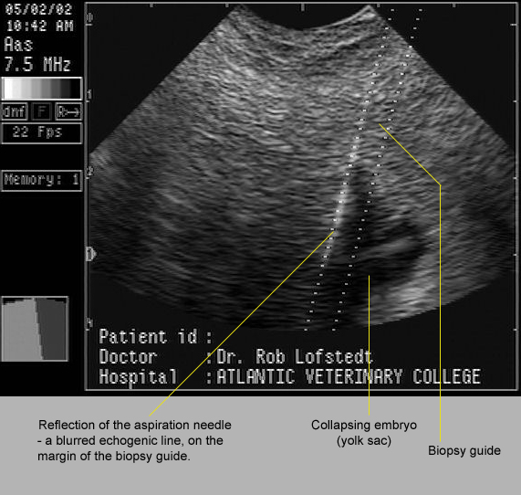
In the image below, 17 day old twins had fixed close to one another within the uterus. One was punctured and drained by ultrasound-guided aspiration through the fornix of the cranial vagina. If one has access to the correct equipment, this is a straightforward procedure.


Image size:571 x 542px

In many cases, co-twins are not seen and pregnancy progresses. This usually leads to one of several outcomes. Some are shown here.

In the image below, a vesicle containing a degenerating structure believed to be an embryo, was seen on the chorion of its 60 day old co-twin. Judging from the size of this vesicle the conceptus was probably about 40 to 45 days old at the time of its death. The age of the surviving co-twin was not recorded but was probably about 65 days. 


Image size: 1569 x 986px

It is possible for a dead co-twin to co-exist with a live fetus until term. Occasionally, the small, mummified fetuses will be found within the placenta of the normal foal. In other cases, the pregnancy may not go to term. This is exemplified by the case illustrated below. The mummified fetus had obviously been dead for some time yet the surviving co-twin appeared to have been normal until shortly before it was aborted. Therefore, the reason for death was not obvious in this case. Indeed, it is tempting to suggest that abortion may have been due to an unrelated problem such as EHV1 infection.


Image size:1218 x 870px Modified from original. Copyright Copyright: Dr Cyril Stephen. Charles Sturt University. NSW.  Au. cyrilstephen@gmail.com

In most cases, the cause for aborted twin pregnancies appears to be related to the presence of its a co-twin. The reason for abortion is still poorly understood but may involved immunological incompatibility, endometrial  sharing and other mechanisms.  

Inset images A, B and C in the amalgam below show abortions that were probably related to the fact that they were twin pregnancies. These cases occurred many years ago, long before the use of ultrasound in stud practice and before the groundbreaking work of Dr O.J.Ginther, a pioneer in our understanding of twinning. Nowadays, with optimal stud management, similar images should be rare. 

In the last image (D) one can see the result of a twin pregnancy that progressed to term. Occasionally normal twins are born but most twins require intensive and expensive neonatal treatment. Even so, many of these foals die or are euthanized after prolonged treatment.


Image size: 1569 x 1989px

Interestingly, it is more common for the smaller neonate to survive than its larger co-twin. In that regard, it has been suggested that the smaller co-twin is under more stress than its co-twin, accelerating fetal maturation.  

It should also be noted that twin pregnancies are also potential causes of dystocia during abortion or during birth at term. In inset C, a "wryneck" fetal malformation in the larger foal complicated that delivery more than otherwise. 

A cautionary note:
Often a mare is presented with premature lactation; a strong indication that abortion is imminent. If the mare has not been under one's care from the time of conception, it is just as well to warn the owner that twins may be causing the abortion. Naturally, an effort should be made to exclude placentitis. Also, the owner's expectation as to the efficiency of EHV1 vaccination should be tempered with reality. However, the possibility of twins should not be discarded even if twin heart beats cannot be discerned on transabdominal ultrasonography on repeated examinations. 


Thursday, February 19, 2015

Umbilical cord length and abortion

Keywords: abortion, equine, umbilical, cord, length

Abnormalities of the umbilical cord have been implicated as causes of abortion in mares. In some cases, a cord abnormality suggests strongly that it has resulted in abortion. In the absence of funisitis (inflammation of the umbilical cord) excessive length and twisting are two suggested causes. Some suggest that 6 or more twists my be abnormal but more twists than that can be seen in normal pregnancies. Sacculation of the urachus and mild contusions of the cord are less convincing as causes of abortion. Some data suggests that umbilical cords are longer in older mares and are in turn, associated with abortion. However, it is difficult to separate the effects of long umbilical cords from those of chronic endometrial fibrosis and other defects associated with aging.

Opinions vary as to how long a cord must be to be considered pathological. Some colleagues consider cords with a total length of more than 80 centimeters (intra- plus extra-amnionic) to be abnormal; others, over 100 cm. Therefore caution must be exercised in implying that the length of the cord has played a role in abortion. Many normal foals are born with cord lengths within the range of 80 to 100 cm. The reason for associating cord length with abortion is remains poorly understood but it may be related to disturbances of blood flow within the cord, especially when they are more than five or six twists in a cord.

In the following image, the two placentas are from two normal foals born on the same day. One has a cord that is 45 cm in length; the other, 92 cm.


Image size: 1500 x 948px

In the following case, the foal was also normal, yet the cord was more than 2 meters long. The extra-amnionic cord alone measured 200cm. With a modest 40cm added for the length of the intra-amnionic cord, the total length would have been approximately 240cm. Interestingly, the umbilical cord was unusually long during the previous foaling but its length was not recorded. It is not known if the sire was the same in both cases.


Image size: 1208 x 1036px. Copyright: Dr Cyril Stephen. Charles Sturt University. NSW.  Au. cyrilstephen@gmail.com


Image size: 1650 x 1409px

In the six month old fetus aove, the intra-amnionic cord alone was 75 cm in length. The extra-amnionic cord was not submitted because it was disposed of when the retained placenta had been expelled. Had that part of the cord been included, the total length would almost certainly been longer than 100cm. Even at term, that would have been considered to be long. Therefore abortion may indeed have been due to its long umbilical cord. In addition, there were 11 twists in the intra amnionic portion of the cord. Finally, the cord showed sacculations of the urachus. In normal foalings these urine-filled sacculations do occur but are not common. Insets show the length of the intra-amnionic cord and urine in a large sacculation.

In the main image, forceps have been inserted into the external orifice of the urachus, where urine drains into the allantois. The extra amnionic cord had been ripped off at this point, its vascular fragments visible adjacent to the forceps.

Note: Excessively short cords may also be abnormal. They have been implicated as a cause of premature rupture during the second stage of foaling and asphyxia

References:

Hong, C.B. et al. 1993  Equine abortion and stillbirth in central Kentucky during 1988 and 1989 foaling seasons. Vet Diagn Invest 5:560-566

Tengelsen, L.A. et al 1997. A 12-year retrospective study of equine abortion in Michigan Vet Diagn Invest 9:303-306

Whitwell. K.E. 1975. Placental pathology: Morphological studies on the normal singleton foal at term. Res. Vet. Sci. 19: 44-55.

Wednesday, February 5, 2014

Ascending placentitis in a Standardbred mare

Keywords: ascending, placentitis, yeast, bacteria abortion

Note: Although is scientifically more accurate to refer to the fetal components of the placenta as the fetal membranes rather than the placenta (the placenta consisting of both fetal and maternal components) common usage will prevail in this discussion.

A pluriparous Standardbred mare aborted a fetus at approximately 9 months of gestation. This would have been her fourth pregnancy. The foal was in good body condition at presentation and although the placenta was abnormal as discussed below, the fetus itself showed no macroscopic abnormalities.

The placenta and fetus were attached i.e. the fetus and its placenta had been expelled as a unit.

Image size: 1500 x 1156px

The crown rump length of the fetus was approx  80 cm and its umbilical cord was 66 long with approximately equal  intra-amnionic and extra-amnionic lengths. The cord was not excessively twisted and showed no macroscopic abnormalities. Also, the mare had been vaccinated repeatedly against EHV1, as is generally recommended.

Once the fetus and its placenta were cleaned and arranged for inspection, severe placentitis was evident in the placenta that had occupied the body of the uterus. The affected area reached from the cervical star to the bases of the placenta that had occupied the uterine horns. As shown below, a strip of normal placenta was present along the right side of the placenta, adjacent to the area of placentitis.

 Image size: 1000 x 667px

A clear line of demarcation was present between the healthy and affected areas. It appeared as though an infectious process had begun in the region of the cervical star and had progress cranially and laterally across the body of the chorion. In the image below, the cervical star is barely visible on the right side of the chorion.

Image size: 975 x 650px

Histology was examined under the yellow ring i.e. an especially thickened, pale and rough area of the affected placenta. This area was completed denuded of villi and disseminated focal, suppurative placentitis was present. This is seen below:

Image size: 1200 x 905px

A higher power view of the neutrophilic infiltration is also shown:

Image size: 1200 x 904px

A closer view of the line of demarcation shows healthy chorion (chorioallantois) on the right and the denuded, abnormal chorion on the left. The area under the yellow ring was sampled for histology 

Image size: 1500 x 1000px

Low power histology of the ringed area is shown below. Normal microcotyledonary placentation can be seen in the sub-image at right. The margin between normal and abnormal tissue is shown in the sub-image on the left. On the extreme left, the villi have been completely destroyed.

Image size: 1200 x 465px

Complete destruction of the villi in the margin are is shown below on high power, again with substantial infiltration of neutrophils:

Image size: 1360 x 1024px

Interestingly, many collections of spore-like bodies (shown under yellow rings) were present in both the areas of destruction.....

Image size: 1360 x 1024px

....and in the adjacent areas where villi were still present (see below).

Image size: 1360 x 1024px

Paint brush petechia were present on the allantoic surface of the amnion (the allanto-amnion). These areas were not examined histologically. The petechia were presumed to be due to a local inflammatory product from the allantochorion. Histology of the fetal adrenals, lungs, kidney and liver was normal.

Image size: 1500 x 1035px

Large vessels present in the amnion (red arrow) were consistent with the immature state of the fetus.

 Image size: 1033 x 703px

In an attempt to demonstrate an etiologic agent, tissue sections of the placenta in the areas of chorionic villus destruction were stained with Periodic acid–Schiff (PAS) and later, with Grocott-Gomori's (or Gömöri) methenamine silver stain (GMS). Both of these stains are commonly used to identify fungi and yeasts, especially PAS which is quicker, cheaper and more convenient stain to use than GMS. 

Using PAS stain, no etiologic agent could be seen. However GMS showed large populations of yeast-like organisms in the affected areas:

 Image size: 1000 x 688px

It was presumed that it was these organisms and smaller forms of the same, that had been identified as "spore-like structures" on the H&E sections discussed earlier in this entry.

Thanks to Dr Peter Sudbery (P.Sudbery@shef.ac.uk) and Dr Mats Troedssen and colleagues at the Gluck institute Ky. for their opinions. These organisms were tentatively identified  as yeasts. According to Dr Sudbery, because of their small size and absence of filamentous forms, they are unlikely to be C. albicans. 

As shown below (with kind permission of Dr Sudbery) filamentous forms of C. albicans are common.

Image size: 960 x 1999px, copyright Dr Peter Sudbery (P.Sudbery@shef.ac.uk)

A MacDonalds gram stain also revealed the presence of large numbers of Gram positive coccobacilli bacteria deep in the chorion. This indicated that these bacteria were probably not contaminants and were most likely participants in the pathogenesis of placentitis. 

Image size: 1000 x 753px

In consultation with a pathologist, it was not possible to say what genus bacteria these were.

Due to the potentially destructive nature of the yeast-like pathogen and the Gram positive bacteria, we have suggested to the mares' owner that an endometrial biopsy should be examined to determine the prognosis for fertility. Also, endometrial cytology should be studied for the continuing presence of pathogens. In the event that the yeast-like organism is still present, a 1% irrigation solution of clotrimazole should be used for treatment. In North America this is compounded for veterinary use (http://www.wedgewoodpetrx.com) and could easily be compounded elsewhere. Antibiotic treatment may also be indicated.

Finally, the cervix of this mare should be examined for tears or any other imperfections that may have contributed to ascending placentitis.

Reference: Sudbery et al. 2004. The distinct morphogenic states of Candida albicans. Trends in microbiology. 12:317-324.


Friday, November 22, 2013

Abortion, eCG and infertility

Keywords: abortion, eCG, infertile

An aborting equine pregnancy.


Image size: 1200 x 835px

This old but unique image shows a pregnancy in the process of aborting at approximately 55 days of gestation, midway through the breeding season.

There is considerable physiological and practical importance attached to this situation. The primary corpus luteum (black arrow) and secondary corpora lutea (in the contralateral ovary and not visible here) had undergone luteolysis. Yet, if this mare had not died, it is unlikely that she would have begun to have estrous cycles again during that breeding season.This is because the endometrial cups (yellow arrows) persist, even after the death of the fetus. The patho-physiological reason for this is unknown and somewhat mysterious. The mystery lies in the fact that the advent of endometrial cups at about 28 to 35 days of gestation is marked by eCG production by the cups and ovulation of follicles; hardly inactive events. However, those same endometrial cups and eCG are associated with ovarian inactivity or unpredictable follicle growth and failure of ovulation if the fetus should die. In most cases therefore, if an equine fetus dies after 35 to 40 days of gestation, the mare cannot be re-bred during the same season. In a minority of mares however, ovarian activity may begin again and a few of these mares will even ovulate and even conceive.

Further mystery is engendered by publications from Argentina, where male pregnancies are aborted after sexing because females are more desirable as polo ponies. Because sexing of the equine fetus is routinely performed at about 60 days of gestation, endometrial cups should be well-formed and functional in these mares. Yet, there are reports of significant numbers returning for breeding in the same breeding season. The reasons for this discrepancy in world-wide findings is not known.

Because cells from the embryo invade the endometrium when endometrial cups are formed they are afforded support from the endometrium and are able to live independently of the fetus. It is only after immunological rejection of the cups at about 120 days of gestation, that mares would be expected to start to have estrous cycles again. However, this usually after the breeding season has ended. Interestingly, in some mares endometrial cups persist for even longer, to well over 200 days and in a few cases to beyond foaling!

With high frequency ultrasound transducers, endometrial cups can sometimes be seen by transrectal ultrasonography. If this is not possible and a mare is anestrus or cycling irregularly, an eCG assay may clarify the situation. However an eCG assay is only useful for a short period after abortion but only soon after abortion (two months at most) because eCG concentrations decrease rapidly after abortion despite the continued presence of endometrial cups.

PS:  First, note the concave shape of the endometrial cups when the image is enlarged; a somewhat lame reason (in the author's opinion) for referring to them as "cups". Perhaps endometrial "plaques" would have been more fitting, especially because may are not even circular but elongated, like slug trails. Secondly,  note the presence of a para-ovarian cyst in this image (encircled by the yellow ring); often thought to be a remnant of the Wolffian (mesonephric) system. However these structures are sometimes shown to be of Mullerian (paramesonephric) origin. There is even confusion in the human literature on that subject.

Selected reference:

Estrade, M.J. et al. 2016 eCG Concentration and subsequent reproductive activity in mares after abortion at day 70. J.Equine Vet.Sci. 42:88-93

Friday, September 20, 2013

Endometrial cup formation


Keywords: equine, mare, eCG, endometrial, cups, uterus, abortion


Image size: 1500 x 1126px

Endometrial cup formation at about 45 days of gestation. The fetus has been removed from its placenta and placed on the endometrial cups as a source of reference.

A section of the endometrial cup/s is indicated here by the green arrows. In this authors opinion, the term "cup" is misleading because these structures are only cupped in cross section but otherwise bear no resemblance to cups. In fact, as is seen here, the cups are often elongated and some cups coalesce with others. They are irregular in shape and vary greatly in size within a single pregnancy.

The cups are actually of fetal origin, producing eCG, formally called pregnant mare serum gonadotropin or PMSG. Equine chorionic gonadotropin production usually starts at about 28 to 35 days of gestation and continues to about 120 days of gestation, sometimes longer and on rare occasions, throughout pregnancy.

The function of eCG is an enigma (2013) because it is associated with multiple ovulation during pregnancy yet cannot induce superovulation in non-pregnant mares. Also, as seen here, most mares have multiple ovulations, forming accessory corpora lutea while eCG is being produced. Yet, a minority have normal pregnancies with no accessory corpora lutea.

A further intriguing finding is that the production of eCG persists after abortion and in most cases usually causes prolonged anestrus. There is no easy and effective method for shortening the life of endometrial cups once they have formed therefore it is advisable to perform therapeutic abortions before day 28, when endometrial cups begin to form. However this situation requires clarification because recent reports on polo ponies challenge these assertions. In polo ponies male pregnancies are often aborted after fetal sexing at about 60 days yet many of those mares have estrous cycles after abortion.

Selected reference:

Estrade, M.J. et al. 2016 eCG Concentration and subsequent reproductive activity in mares after abortion at day 70. J.Equine Vet.Sci. 42:88-93