EQUINE IMAGES

How to use these images


IMPORTANT: To see an enlarged image, click on any image you see in LORI. Then, RIGHT click on the enlarged image to save it at its full size.


Showing posts with label infertility. Show all posts

Monday, March 28, 2016

Treatment of retained placentas

Keywords: equine, placenta, retained, Burn's, infertility

The incidence of retained placenta in mares has been reported to lie between 2 and 11 % of all foalings in most breeds. In other breeds, especially Friesians and draft horses, it can be far higher. Unlike the situation in cattle, retained placenta in mares is a potentially life-threatening disease.

If the placenta is still attached three or more hours after foaling, it should be considered retained. Although it has been stated (like the situation in cattle) that uterine atony does not contribute to retained placentas, the vast majority of retained placentas in mares are expelled after one or more injections of oxytocin; certainly not the case in cattle. In the author's experience, simple treatment with 10 iu of oxytocin i.m. every 30 minutes will dislodge the vast majority of retained placentas within five or six treatments. Treatment with continuous I.V. drips has been described but it is not clear if that method warrants its extra expense and inconvenience. As a nonapeptide, oxytocin is a small and stable molecule and is very rapidly absorbed after intramuscular injection. With little potential for abuse, it can be left with responsible horse owners together instructions for treating retained placentas. Placentas that are still retained after six treatments should probably receive intensive veterinary care.

A novel, effective, and apparently safe method of treating retained placenta in mares was described by Meijer et al in 2015. Briefly, this involves infusing water into any major umbilical blood vessel for several minutes until the placenta becomes dislodged. It has yet to become widely adopted.

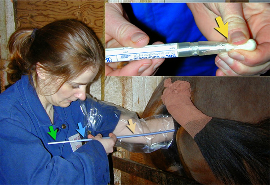
An alternative treatment is to inflate the chorioallantois with fluid to loosen its attachment to the endometrium and presumably, to stimulate myometrial contraction. This is colloquially known as the Burns technique. The author has not found the Burn's technique to be reliable but has little experience with the procedure.

The collage of images below shows the typical appearance of a retained placenta and the application of the Burns technique in a mare. About 10 to 12 liters of saline are infused into the intact chorioallantois and the site of infusion is ligated with umbilical tape to retain the saline.


Image size: 1572 x 2046px

If the placenta has not been expelled within 30 minutes, the ligation is removed and the fluid is allowed to drain. Conventional care is then adopted. This care includes oxytocin, tetanus prophylaxis, antibiotics, non-steroidal anti-inflammatory drugs, and in the view of some, vasodilators to prevent laminitis.

Manual extraction of the placenta remains a contentious subject. In North America, the general belief is that the placenta should not be manually removed. Instead, a mare should be treated systemically (as described above) until her placenta has been expelled. This may take two or three days to occur. However, in Europe it is common to remove placentas manually if they have been retained for longer than three hours. Indeed in the Netherlands, it is actually considered to be malpractice if the placenta is not removed manually when it has been retained for longer than six hours post foaling. Interestingly, at least two peer reviewed studies have shown than manual removal of fetal membranes did not affect reproductive performance.


Selected references:

Canisso, I.F. et al 2013. A clinical approach to the diagnosis and treatment of retained fetal membranes with an emphasis placed on the critically ill mare. J.Equine.Vet Sci. 33: 570-579

Cuervo-Arango, J.et al. 2009 The Effect of manual removal of placenta immediately after foaling on subsequent fertility parameters in the mare. J. Equine.Vet Sci. 29:771-774

Gibbens, D. et al. 1972. The circulating levels of oxytocin following intravenous and intramuscular administration of syntometrine. British J. Ob.Gyn. 79:644-646

McKinnon, A.O. et al Eds. 2011, Equine reproduction. Second ed. Chapter 260. Wiley-Blackwell ISBN: 978-0-8138-1971-6

Meijer, M et al. 2015. How to use umbilical vessel water infusion to treat retained fetal membranes in mares. Proceedings AAEP 61:478-484

Paccamonti, D.L. et al 1999. PGFM response to exogenous oxytocin and determination of  the half-life of  oxytocin in nonpregnant mares. Equine Vet J. 31:285-288

Provencher, R. et al. 1998. Retained fetal membranes in the mare: A retrospective study
Can Vet J. 29: 903–910.

Rapacz, A. et al. 2012. Retained fetal membranes in heavy draft mares associated with histological abnormalities. J. Eq. Vet. Sci. 32: 38-44

Sevinga et al 2002. Reproductive performance of Friesian mares after retained placenta and manual removal of the placenta. Theriogenology.57: 923–930

Sevinga, M. et al 2002. Serum calcium and magnesium concentrations and the use of a calcium-magnesium borogluconate solution in the treatment of Friesian mares with retained placenta
Theriogenology 57: 941–947


Monday, March 2, 2015

Twins and infertility

Keywords: equine, infertility, twins, ovulation, abortion.

Note: the term conceptus is used here by the author, in preference to embryo. In the author's mind, conceptus refers to the embryo together with its membranes while embryo refers to the future fetus. Also, because of the importance of twinning in equine reproduction, the author has included more text than usual in this entry.

Twin preovulatory follicles seen on ultrasound.


Image size: 900 x 694px

Conventional wisdom once dictated that mares should not be bred when twin preovulatory follicles were present. This was based on the fact that twins are a common cause of abortion in mares. With the increase in knowledge that came with common use of ultrasonography, this philosophy has changed. Today, it would be considered poor management to miss this breeding opportunity. Indeed, if mares where not to be bred whenever twin follicles were present, perhaps 15 to 20% of all breeding opportunities would be lost. This is especially the case in breeds such Thoroughbreds and Standardbreds where twin ovulations are common. It is also significant in any breed with the approach of the summer solstice because day length and ovulation rate are positively correlated.

Current philosophy dictates that mares should always be bred when twin follicles are present. Indeed, even when twin pregnancies are not discovered, the overall singlet pregnancy rate is higher when this approach is used.


Notes on the timing of pregnancy diagnosis:

When two conceptions occur during a single estrous period, one conceptus will invariably be older and larger than the other conceptus when pregnancy diagnosis is performed. This is important to bear in mind. Although not common, twin ovulations can be separated by several days, making it possible to miss a younger conceptus when routine pregnancy diagnosis is performed at 14 to 15 days after ovulation. 

Newer, high resolution ultrasound units make it possible to see pregnancies at 12 days or even earlier. If there a second, younger conceptus in the uterus at this time, it may not be seen. However, if pregnancy diagnosis is delayed to 14 days, it is very likely that both conceptuses will be seen. After 16 days, embryo movement within the uterus will have ceased and the opportunity to crush one embryo without harming the other will have diminished significantly. Therefore pregnancy diagnosis should be scheduled at 14 to 16 days after the time that the first of two intact (preovulatory) follicles was last seen. This is explained below:

On many stud farms, visits are made on Mondays, Wednesdays and Fridays. If the time of ovulation is taken as the time a corpus luteum is first seen on ultrasonography, it can amount to a critical error in timing of pregnancy diagnosis. This is because the actual time of ovulation could have been two or even three days earlier, immediately after one last saw an intact follicle.

Finally, two large follicles may be present yet, with the vagaries of ultrasonography and work pressure, it is not rare for one to be under the impression that there is only a single pre-ovulatory follicle. Therefore one should always assume that twins may be present unless proven otherwise!


Taken 14 to 15 days after the last record of an intact follicle, the images below show how quickly embryos can migrate within the uterus before the time of fixation. This is used to one's advantage when crushing one of the embryos (usually the smaller of the two).


Image size: 1800 x 1369px

Movement through the uterus is a product of myometrial contraction i.e. even inanimate objects will move quickly within a mare's uterus. This is easily demonstrated by inserting small, sterilized fluid- filled "balloon twins" into the uterus via the cervix.


Image size: 1308 x 905px

Movement of embryos is thought to be important in the recognition of pregnancy but stops abruptly at about 16 days after ovulation.

In practical terms, it is very important for the veterinarian to examine every part of the uterine horns and body before excluding the possibility of twins.  Sometimes one of the twins may be present in a uterine horn while its co-twin is found in the uterine body, just cranial to the cervix.  It is quite easy to miss a co-twin in the uterine body if it is slightly off-center. Care must be taken to ensure that the specular reflection of the endometrial surface is visible as a thin echogenic line along the entire length of the uterine body from cervix to bifurcation. An illustration of this echogenic line can be seen in first image of this LORI entry

As mentioned, it is common to crush one co-twin to prevent abortion of both fetuses later in gestation. After tranquilization and rectal relaxation (n-butylscopolamine) as needed, the author locates the co-twin to be crushed and withdraws the transducer. The conceptus is then crushed immediately (before it is moved by the myometrium) as shown below. If the crush has been successful, little or no fluid will remain visible. Occasionally, several attempts at crushing may be required.


Image size: 800 x 552px

If there is concern that attempts at crushing have released enough prostaglandin to cause luteolysis, altrenogest (Regumate™) should be administered until the accessory corpora lutea have formed and the serum progesterone concentration has risen to well over 2ng/ml. Fortunately, altrenogest does not cross-react with most progesterone assays, so endogenous progesterone concentrations can be monitored until it is safe to wean the mare off altrenogest treatment.  

If one fails to crush a co-twin or if pregnancy diagnosis has been delayed beyond 16 days, other methods of twin reduction must be adopted. If a co-twin is crushed after this time, it is likely that both conceptuses will die; the reasons for that being beyond the scope of this discussion. Twin reduction using trans-vaginal ultrasound guided aspiration can be successful in these cases and should be attempted in favor of crushing if the critical day 16 has passed.

In the image below, 17 day old twins had fixed close to one another within the uterus. One was punctured and drained by ultrasound-guided aspiration through the fornix of the cranial vagina. If one has access to the correct equipment, this is a straightforward procedure.


Image size:571 x 542px

In many cases, co-twins are not seen and pregnancy progresses. This usually leads to one of several outcomes. Some are shown here.

In the image below, a vesicle containing a degenerating structure believed to be an embryo, was seen on the chorion of its 60 day old co-twin. Judging from the size of this vesicle the conceptus was probably about 40 to 45 days old at the time of its death. The age of the surviving co-twin was not recorded but was probably about 65 days. 


Image size: 1569 x 986px

It is possible for a dead co-twin to co-exist with a live fetus until term. Occasionally, the small, mummified fetuses will be found within the placenta of the normal foal. In other cases, the pregnancy may not go to term. This is exemplified by the case illustrated below. The mummified fetus had obviously been dead for some time yet the surviving co-twin appeared to have been normal until shortly before it was aborted. Therefore, the reason for death was not obvious in this case. Indeed, it is tempting to suggest that abortion may have been due to an unrelated problem such as EHV1 infection.


Image size:1218 x 870px Modified from original. Copyright Copyright: Dr Cyril Stephen. Charles Sturt University. NSW.  Au. cyrilstephen@gmail.com

In most cases, the cause for aborted twin pregnancies appears to be related to the presence of its a co-twin. The reason for abortion is still poorly understood but may involved immunological incompatibility, endometrial  sharing and other mechanisms.  

Inset images A, B and C in the amalgam below show abortions that were probably related to the fact that they were twin pregnancies. These cases occurred many years ago, long before the use of ultrasound in stud practice and before the groundbreaking work of Dr O.J.Ginther, a pioneer in our understanding of twinning. Nowadays, with optimal stud management, similar images should be rare. 

In the last image (D) one can see the result of a twin pregnancy that progressed to term. Occasionally normal twins are born but most twins require intensive and expensive neonatal treatment. Even so, many of these foals die or are euthanized after prolonged treatment.


Image size: 1569 x 1989px

Interestingly, it is more common for the smaller neonate to survive than its larger co-twin. In that regard, it has been suggested that the smaller co-twin is under more stress than its co-twin, accelerating fetal maturation.  

It should also be noted that twin pregnancies are also potential causes of dystocia during abortion or during birth at term. In inset C, a "wryneck" fetal malformation in the larger foal complicated that delivery more than otherwise. 

A cautionary note:
Often a mare is presented with premature lactation; a strong indication that abortion is imminent. If the mare has not been under one's care from the time of conception, it is just as well to warn the owner that twins may be causing the abortion. Naturally, an effort should be made to exclude placentitis. Also, the owner's expectation as to the efficiency of EHV1 vaccination should be tempered with reality. However, the possibility of twins should not be discarded even if twin heart beats cannot be discerned on transabdominal ultrasonography on repeated examinations. 


Wednesday, February 25, 2015

Streptococcus equi zooepidemicus


Keywords: Streptococcus equi var. zooepidemicus, infertility, equine

This image shows a culture of Streptococcus equi var. zooepidemicus on blood agar. The author's record is devoid of details for this image but it was probably isolated from a mare with endometritis because it is a common genital pathogen in mares.

Streptococcus equi var. zooepidemicus is also a common commensal bacterium, easily isolated from normal horses. This is important to bear in mind when cultures are positive for Streptococcus zooepidemicus because contamination from the perineal area will often produce false positive cultures.


Image size: 1664 x 1440px

The image shows beta hemolysis (complete hemolysis) around the colonies of bacteria. This is due to an exotoxin produced by many Streptococcal species. Some of those species cause partial hemolysis (alpha hemolysis) as well, but not Streptococcus equi var. zooepidemicus

Note: It is common for clinicians to refer Streptococcus equi var. zooepidemicus by its short form, Streptococcus zooepidemicus or even more colloquially,  "Strep zoo", forgetting perhaps that this bacterium is one of two subspecies of Streptococcus equi . The other is Streptococcus equi  var. equi , the well known cause of Strangles in horses. Streptococcus equi var. zooepidemicus is a gram-positive coccoid bacterium occurring in pairs or long chains on stained preparations. It is usually sensitive to penicillin and its derivatives.

Tuesday, February 24, 2015

Streptococcus equi zooepidemicus


Keywords: Streptococcus equi var. zooepidemicus, infertility, equine

This image shows a culture of Streptococcus equi var. zooepidemicus on blood agar. The author's record is devoid of details for this image but it was probably isolated from a mare with endometritis because it is a common genital pathogen in mares.

Streptococcus zooepidemicus is also a common commensal bacterium, easily isolated from normal horses. This is important to bear in mind when cultures are positive for Streptococcus zooepidemicus because contamination from the perineal area will often produce false positive cultures.


Image size: 1664 x 1440px

The image shows beta hemolysis (complete hemolysis) around the colonies of bacteria. This is due to an exotoxin produced by many Streptococcal species. Some of those species cause partial hemolysis (alpha hemolysis) as well, but not Streptococcus equi var. zooepidemicus 

Note: It is common for clinicians to refer Streptococcus equi var. zooepidemicus by its short form, Streptococcus  zooepidemicus, forgetting perhaps that this bacterium is one of two subspecies of Streptococcus equi . The other is Streptococcus equi  var. equi , the well known cause of Strangles in horses. Streptococcus equi var. zooepidemicus is a gram-positive coccoid bacterium occurring in pairs or long chains on stained preparations. It is usually sensitive to penicillin and its derivatives.

Tuesday, December 9, 2014

Foaling injuries and pneumometra (wind sucking)

Keywords: pneumometra, wind-sucking, equine,injury, infertility

Injuries from foaling or other causes may result in mild to severe distortion of the vulvar lips after healing.  The result is essentially the same as if a mare had congenitally poor conformation i.e. affected mares becomes "wind suckers” and pneumometra develops.


Image size: 1800 x 1622px

The cause of the injury in the mare at upper left was never determined but it was not due to foaling. This contributed to wind sucking. In the image below that, mild distortion of the left vulva lip was indeed a result of foaling. Surprisingly however, this did not appear to cause wind sucking.  When Caslick’s operations are not reversed prior to foaling, similar injuries can occur.

When pneumometra occurs, it is occasionally visible using transrectal ultrasonography. For example, in images A and B, (a separate case from the mares pictured at left) bubbles of air can be seen as highly echogenic structures in uterine cross-sections. This can cause long term infertility as discussed elsewhere in LORI.


Saturday, May 24, 2014

Fetal death, eCG; the MIP test

Keywords equine, MIP, endometrial cups, infertility

Endometrial cups usually persist if fetal death occurs after 35 to 40 days of gestation. For unknown reasons, persisting production of eCG often causes failure to return to cyclicity or protracted periods of irregular estrous cycles. Even when a live foal is produced after an apparently normal pregnancy, endometrial cups and eCG production may persist, sometimes causing disruption of "foal heat" ovulations.

Endometrial cups are difficult to see using ultrasonography and one may not possess an endoscope. Therefore it may be necessary to rely on endocrinological tests to ascertain if endometrial cups are still present. It is most common to measure eCG by radioimmunoassay or ELISA  but one may still find this old test in use:


Image size 1180 x 414px

It is a hemagglutination inhibition test, also knows and the mare immunopregnancy (MIP) test, a qualitative assay for eCG.  In this illustration, the set of tubes on the right side have produced a positive test result. Endometrial cups were present in this mare. They can be seen on the endoscopic view at extreme right. It should be emphasized that the primary reason for marketing this test was to detect pregnancy, not to diagnose the persistence of endometrial cups. It can be seen however, that the MIP test was prone to false positives because a mare could still test positive for pregnancy after fetal death had occurred.

The image on the left hand side shows a negative test result.

Routine culture, sensitivity and cytology 

Keywords: diagnostic, techniques, equine, infertility, stud.

During routine breeding soundness evaluations or when fluid is seen in the uterus on ultrasonography prior to breeding, both culture and cytology are commonly performed.

The image below shows routine endometrial culture in one practice. The blue, green and light orange arrows indicate the double guarded nature of the culture instrument. The yellow arrow at top right shows how the bacterial culture swab is transferred to a special transport medium immediately after culture. Common culturing instruments for mares do not contain transport media and are inclined to dry out before they can be plated for culture.


Image size: 1127 x 772px

In the three sub-images shown here, both culture and sensitivity are demonstrated.   Plate 1 shows a pure culture of  a Coliform species Plate 2 shows beta hemolysis (alpha hemolysis is less obvious) caused by a culture of Streptococcus zooepidemicus, a common commensal and pathogen in mares. Plate 3 is an antibiogram, showing clear growth inhibition by two antibiotic discs and little inhibition by the others.


Image size: 981 x 691px

Immediately after withdrawal of the culture section of the guarded sampling system, a cytology sample is collected using a PAP cervical cytology brush. The cytology brush (wedged into the end of a standard infusion pipette) is inserted into the system, which is still  in the uterus. The brush tip, seen below, collects an excellent sample of endometrial cells and the procedure only requires a single entry into the uterus.


Image size 1200 x 800px

These brushes can be obtained in large quantities and low cost. The brush base is inserted into the end of the plastic infusion pipette and sterilized before use. In our experience, the cytology samples obtained in this manner are far superior to those obtained with a rayon swab. The brush tip is rolled (not smeared) onto two glass slides. One slide is stained with Diff-Quick (modified Wrights-Giemsa) and examined immediately. The other slide is retained for Grams staining in the event that inflammation is seen. A cytology specimen collected from the uterus of a mare is shown here.


Image size: 999 x 710px

The black arrow shows normal epithelial cells from the endometrium; the length of their cytoplasmic "tail" indicating that this mare was in estrus at the time of sampling. The large number of  lymphocytes (green ring) is very unusual its significance in this mare was not determined. Although lymphocytes are often seen in the lamina propria in mares with chronic endometritis, they are seldom seen in the uterine lumen. A few neutrophils in this preparation (red ring) indicate that there is an acute component to this inflammation as well.

Interestingly, in most mares with uterine fluid (routinely examined in one large stud practice) signs of uterine inflammation are very seldom seen and occasionally samples of the clear fluid are collected and incubated with freshly collected sperm. Sperm survival in these samples is superior to survival in non-extended semen. Therefore the significance of intrauterine fluid itself  is questionable, perhaps reflecting abnormal uterine drainage rather than an intrinsic hazard.

In routine examination of the endometrium, culture is always performed first, assuming that all other procedures have the potential to contaminate the uterus and produce false positive culture results.


Monday, October 21, 2013

Endometrial biopsy cytology


Keywords: biopsy, equine, endometrium, uterus, infertility


Image size: 990 x 630px

This image shows margination of neutrophils (PMN) during estrus. This is a common phenomenon seen in endometrial biopsies especially but not exclusively during standing heat. Neutrophils attach to the endothelium of the capillaries in the lamina propria and migrate into the stroma. This is thought to augment uterine defenses during breeding. Also seen in this image are some marginating eosiniphils (E). They are commonly seen in the stroma of equine endometrial biopsies and although they have been associated with the "wind-sucking" condition, this is poorly substantiated.

The capillary containing these cells is surrounded by endometrial glands with lumens that are not obvious.

The image below shows the typical appearance of lumen epithelial cells during estrus. They are significantly taller than the same cells epithelial cells during diestrus (the luteal phase). Also note the cilia. They are dispersed intermittently across the surface of the epithelium. On a scanning electron micrograph they appear like tufts of  grass on a barren lawn. Between the "tufts of grass" are the intermittent openings of endometrial glands.


Image size: 990 x 630px

Hemosiderin is commonly found in macrophages in endometrial biopsies. Evidence of this is shown below. The presence of hemosiderin, which usually remains in the endometrium for long periods of time, usually reveals that a mare has experienced intrauterine hemorrhage sometime in her life. Although there is practically no overt hemorrhage from the endometrium during or after foaling, mild hemorrhage is likely. Therefore endometrial hemosiderosis probably indicates that a mare has had one or more foals in her lifetime.


Image size: 990 x 603px

Plasmacytes  and lymphocytes are common "janitorial cells" in the lamina propria, especially the stratum compactum as shown below. On occasion, even highly experienced pathologists may have difficulty distinguishing between these two types of cells. They provide local immunity in the endometrium and unless they are present in large numbers should not be interpreted as an indication of chronic endometritis.

Image size: 990 x 636px

Saturday, October 19, 2013

A uterine biopsy

Keywords: uterine, uterus, biopsy, infertility, punch

Endometrial biopsies are important in diagnosing endometritis and other causes of infertility in mares.


.
Image size: 990 x 1242px

The jaws of a biopsy instrument are shown here on the dorsal endometrial surface where biopsies on normally taken. This is because the biopsy instrument is placed in the uterus and the dorsal endometrium is pushed down (per rectum) into the jaws of the punch. Sometimes this is referred to as a "guided" biopsy technique. It is far superior to a "blind" biopsy technique where the endometrial biopsy punch is inserted through the cervix and its jaws are closed in the hope of obtaining a satisfactory biopsy.  This author has read endometrial biopsies for a diagnostic laboratory for many years and most of the unsatisfactory samples were taken using the "blind" technique. In fact, samples of the cervix have often been submitted instead of endometrial biopsies.

When taking a biopsy, It is safer to feed the endometrium into the side of the jaws as shown here rather than into the front of the jaws. A large, deep and penetrating biopsy can result in the latter case. This instrument is made by the Pilling company, USA.

The inset shows the biopsy itself being deposited into a container of Bouin's fluid which is 80% picric acid and 20% formalin.  This is a so-called "hard fixative" suitable for genital and embryonic tissue because the histological architecture is not destroyed when the large amounts of water in these tissues are extracted during processing.  However, the use of Bouin's fluid is by no means mandatory because ordinary formalin also provides satisfactory results.