Cervical cerclage for cervical tears
Keywords: Shirodkar, mares, tear, cervix.
Cervical tearing is an occasional complication of foaling.

Image size: 235 x 236px
If the tear is mild, fertility may be normal. However, when complete cervical closure is impossible, fertility is likely to be compromised. It is often impossible to predict how fertility will be affected unless a cervical tear is very severe because pregnancy can be maintained to term in mares that have obvious cervical tears.
In women with compromised cervices, Shirodkar-McDonald, Wurm or Hefner sutures are used. The image below shows a Shirodkar suture pattern. It is a continuous suture pattern with four bites and no entry into the cervical canal.

Image size: 865 x 481px
The Wurm pattern only consists of two mattress sutures (one at right angles to the other) and it does enter the cervical canal. Therefore the importance of avoiding the cervical canal is not clear. A method of cervical cerclage similar to the Shirodkar-McDonald is described on page 2563 of Equine Reproduction. Second edition. Ed. McKinnon, A.O. et al. Wiley-Blackwell. ISBN 978-0-8138-1971-6.
An alternate technique is shown below. This approach may be easier to apply than others and has the advantage that it can be loosened and tightened as required for re-insemination, abortion or delivery. As this technique is in the process of being developed, the author seeks feedback from equine practitioners who may try the technique or modifications thereof.
Initially, the use of a circumferential suture pattern (below) was investigated but even in post-mortem specimens, the author found this suture difficult to place without special instrumentation to stabilize the cervix and pull it caudally.

Image size: 806 x 710px
In the last iteration, a simple continuous suture consisting of only a single loop was used (see below). Placement of an infusion pipette in the cervical canal helped to stabilize the cervix and served as landmark for placing the sutures. The author found this easier to place than the Shirodkar-type suture.

Image size: 1070 x 1635px
Using a section of Silastic TM or similar tubing, a high-friction brake was created as a substitute for a knot. The tubing used had an outside diameter of approximately 4 mm and suture material was passed though the tubing as shown above. After the first pass, each successive pass was at approximately 90 degrees to the preceding pass.
Image size: 806 x 710px
In the last iteration, a simple continuous suture consisting of only a single loop was used (see below). Placement of an infusion pipette in the cervical canal helped to stabilize the cervix and served as landmark for placing the sutures. The author found this easier to place than the Shirodkar-type suture.
Image size: 1070 x 1635px
Using a section of Silastic TM or similar tubing, a high-friction brake was created as a substitute for a knot. The tubing used had an outside diameter of approximately 4 mm and suture material was passed though the tubing as shown above. After the first pass, each successive pass was at approximately 90 degrees to the preceding pass.
Once the knot substitute had been made, a simple suture (see above) was been placed blindly across the cervix. The suture was pulled tight around the cervix then the tubing was pushed down to the cervical fornix to close the suture.
_______________
A step-by-step series of images follows, showing placement of a cervical suture in a Standardbred mare. In that case, conventional surgical repair of the cervix had already been attempted and as is often the case, it was unsuccessful.
1. The mare is examined for the presence of a preovulatory follicle and inseminated.

2. After confirmation of ovulation, the mare is given 10mg of detomidine and 10mg of butorphanol intravenously for restraint and analgesia .

Image size: 410 x 491px
3. The surgical pack and sterilized SilasticTM tubing is shown here. The length and diameter of the tubing
used can be appreciated from the image.

Image size: 498 x 305px
4. Hands are gloved and arms surgically scrubbed to elbow height.
5. The suture material is fastened securely to one end of the SilasticTM tubing using a standard surgical knot.
MersileneTM or a similar non-absorbent braided suture can be used. Although capillary action is a drawback with braided suture materials, the tubing is able to grip braided suture more securely than if monofilament suture material. When the Shirodkar suture is used in women, braided suture material is used in the majority of cases.

Image size: 695 x 468px

Image size: 526 x 489px
7. A small amount of lubrication is applied to the hand to facilitate entry into the vagina; excessive lubrication causes the needle to be too slippery to hold.

Image size: 340 x 599px
8. Holding the needle in one's hand (larger needles are easier to handle than smaller needles) the suture is placed through the dorsal and ventral aspects of the cervix, on either side of the pipette.

Image size: 426 x 378px
Caution: It is easy to place the sutures too close to the cervical canal, allowing gaps from the cervical tear to remain open, peripheral to the suture. For this reason, sutures should be placed as far from the cervical canal as possible.
Image size: 426 x 378px
Caution: It is easy to place the sutures too close to the cervical canal, allowing gaps from the cervical tear to remain open, peripheral to the suture. For this reason, sutures should be placed as far from the cervical canal as possible.
9. After the needle is placed though the cervix, it is pulled caudally, beyond the level of the vulva lips. Using the pattern shown earlier, the suture is passed through the SilasticTM tubing. This is also shown below.


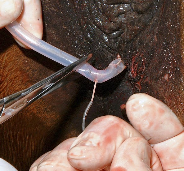
Image size: 376 x 467px
Pass # 1
Image size: 376 x 467px
Pass # 2
Image size: 593 x 553px
Pass # 3
Once the suture has been properly threaded through the tubing, excess tubing is cut off as shown above.
10. Holding the needle in one hand, the tubing is moved down the suture material by sliding it towards the cervix. Because the tubing grips the suture effectively, sliding the tube towards the cervix requires patience.

Image size: 426 x 378px
Caution: Care should be taken to perform this step slowly to avoid the cutting action of the suture material on the cervix.
Image size: 426 x 378px
Caution: Care should be taken to perform this step slowly to avoid the cutting action of the suture material on the cervix.
11. Once the cervix has been closed snugly (but not with excessive tension) excess suture is removed at the vulva lips.

Image size: 473 x 450px
Enough suture is left in the vagina in the event that re-insemination is required. That being the case, a gloved finger is inserted into the cervical canal to loosen the suture. The mare is then re-inseminated.
To re-close the cervix a Caslicks speculum (which has open sides) is used to locate the end of the suture material lying the vagina. It is grasped with a bowel clamp and gradually pulled towards the vulva lips while the tubing is again slid towards the cervix, affording its re-closure.
12. To avoid transluminal adhesions after protracted vaginal interference, the vagina is coated with an oil-based mastitis antibiotic ointment (as is done after cases of protracted obstetrics in mares).

Image size: 442 x 316px
13. To afford better protection of the vagina than otherwise, a temporary Caslick's suture (using 35w steel staples) should probably be used between estrous periods until pregnancy is confirmed. The suture can be converted to a more permanent Caslick's closure when pregnancy becomes established.
Notes
The time for elective release of the suture is under consideration. In that context, it is proposed that impending parturition be predicted by using combined milk electrolyte scores. The Caslick's closure would be opened followed by cervical opening. The cervix would be opened by drawing out the tubing from the vagina and removing the suture from the cervix.
Anticipated problems may include.
1. Fistula formation along the suture tracts.
The author has only used this surgery in two clinical cases. In both cases, outcomes were lost to client/horse movement. Therefore the value of the technique is unknown. In that context, the author (lofstedt@upei.ca) would appreciate suggestions and feedback from equine practitioners who use this technique.
Anticipated problems may include.
1. Fistula formation along the suture tracts.
2. Vaginitis due to the presence of the suture tail in the vagina; a necessity for the entire pregnancy.
3. Spreading the cervical suture in advanced pregnancy due to intrauterine pressure, causing ascending placentitis.
3. Spreading the cervical suture in advanced pregnancy due to intrauterine pressure, causing ascending placentitis.
The author has only used this surgery in two clinical cases. In both cases, outcomes were lost to client/horse movement. Therefore the value of the technique is unknown. In that context, the author (lofstedt@upei.ca) would appreciate suggestions and feedback from equine practitioners who use this technique.