The uterine tube of a mare.
Keywords: uterine tube, oviduct, fallopian tube, equine,
mare, embryo
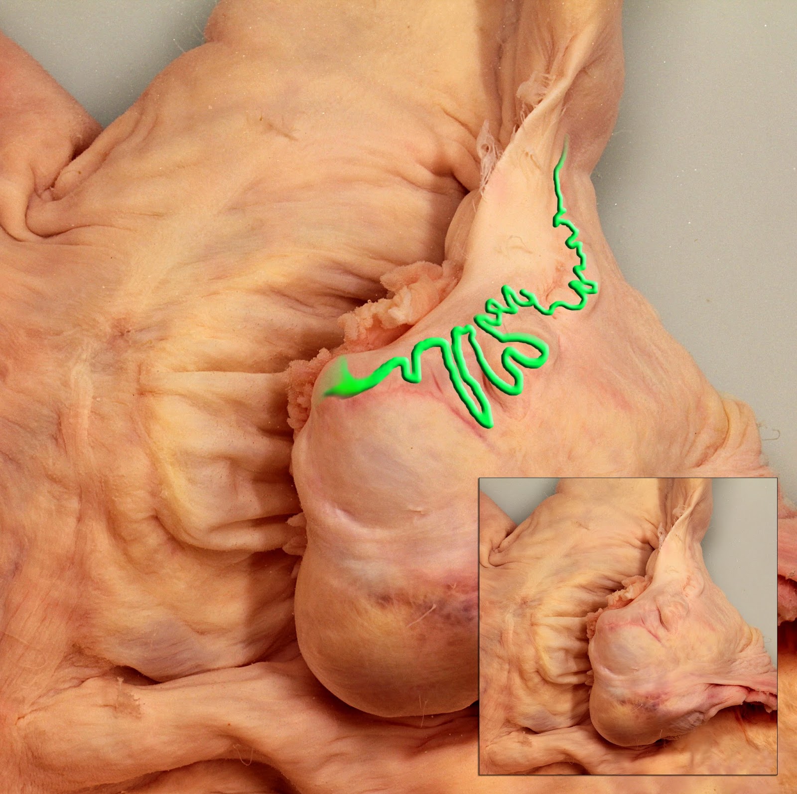
Image size: 3246 x 3234px
The normal orientation of the ovulation fossa in the ovary is ventral-to-lateral. Therefore this is the dorsal aspect of the mesopsalpinx tube of a mare, showing the tortuous passage of the ampulla from the infundibulum (at left) to the isthmus and utero-tubal junction at right. It has been hi-lited in green in the major image. The natural state is shown in the inset.
The normal orientation of the ovulation fossa in the ovary is ventral-to-lateral. Therefore this is the dorsal aspect of the mesopsalpinx tube of a mare, showing the tortuous passage of the ampulla from the infundibulum (at left) to the isthmus and utero-tubal junction at right. It has been hi-lited in green in the major image. The natural state is shown in the inset.
The uterine tube is a fascinating structure, especially in
mares.
In a mare, a five-day-old morula secretes prostaglandin E2
which stimulates the uterine tube (oviduct) to transport it into the uterus.
This is important because ostensibly, only fertilized oocytes (embryos) reach
the uterus in mares. Others are usually retained in the region of the
ampullary-ithmic junction. On occasion however, unfertilized oocytes do reach
the uterus, probably assisted by the simultaneous passage of fertilized oocytes
(embryos). Interestingly, the oocytes from several estrous cycles appear to
accumulate in the uterine tubes because up to five oocytes have been seen in
the uterine tubes, in various stages of health or degeneration. This was
elegantly described and illustrated by
Dr Peter Flood and colleagues (1979. J. Reprod. Fert. 57: 291-294)
Mares are not unique in this regard, long tonged bats,
hamsters and undoubtedly other mammals share this physiology but their embryos
produce signalling hormones other than prostaglandin E2
Most oocytes of domestic mammals (including those of cows)
reach the uterus on the fourth day of ovulation or shortly before that. In pigs
they reach the uterus even earlier, in just under three days. In mares however,
embryos only enter the uterus between 5 and 6 days after ovulation.