Routine culture, sensitivity and cytology
Keywords: diagnostic, techniques, equine, infertility, stud.During routine breeding soundness evaluations or when fluid is seen in the uterus on ultrasonography prior to breeding, both culture and cytology are commonly performed.
The image below shows routine endometrial culture in one practice. The blue, green and light orange arrows indicate the double guarded nature of the culture instrument. The yellow arrow at top right shows how the bacterial culture swab is transferred to a special transport medium immediately after culture. Common culturing instruments for mares do not contain transport media and are inclined to dry out before they can be plated for culture.
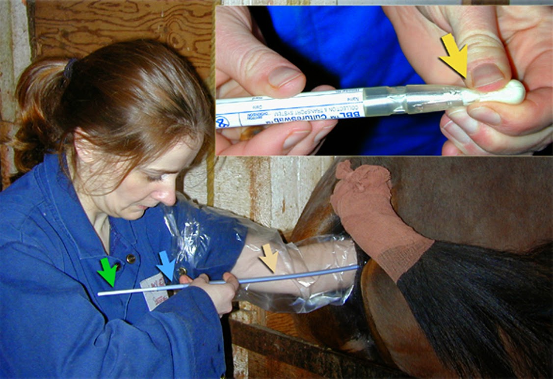
Image size: 1127 x 772px
In the three sub-images shown here, both culture and sensitivity are demonstrated. Plate 1 shows a pure culture of a Coliform species. Plate 2 shows beta hemolysis (alpha hemolysis is less obvious) caused by a culture of Streptococcus zooepidemicus, a common commensal and pathogen in mares. Plate 3 is an antibiogram, showing clear growth inhibition by two antibiotic discs and little inhibition by the others.

Image size: 981 x 691px
Immediately after withdrawal of the culture section of the guarded sampling system, a cytology sample is collected using a PAP cervical cytology brush. The cytology brush (wedged into the end of a standard infusion pipette) is inserted into the system, which is still in the uterus. The brush tip, seen below, collects an excellent sample of endometrial cells and the procedure only requires a single entry into the uterus.

Image size 1200 x 800px
These brushes can be obtained in large quantities and low cost. The brush base is inserted into the end of the plastic infusion pipette and sterilized before use. In our experience, the cytology samples obtained in this manner are far superior to those obtained with a rayon swab. The brush tip is rolled (not smeared) onto two glass slides. One slide is stained with Diff-Quick (modified Wrights-Giemsa) and examined immediately. The other slide is retained for Grams staining in the event that inflammation is seen. A cytology specimen collected from the uterus of a mare is shown here.

Image size: 999 x 710px
The black arrow shows normal epithelial cells from the endometrium; the length of their cytoplasmic "tail" indicating that this mare was in estrus at the time of sampling. The large number of lymphocytes (green ring) is very unusual its significance in this mare was not determined. Although lymphocytes are often seen in the lamina propria in mares with chronic endometritis, they are seldom seen in the uterine lumen. A few neutrophils in this preparation (red ring) indicate that there is an acute component to this inflammation as well.
Interestingly, in most mares with uterine fluid (routinely examined in one large stud practice) signs of uterine inflammation are very seldom seen and occasionally samples of the clear fluid are collected and incubated with freshly collected sperm. Sperm survival in these samples is superior to survival in non-extended semen. Therefore the significance of intrauterine fluid itself is questionable, perhaps reflecting abnormal uterine drainage rather than an intrinsic hazard.
In routine examination of the endometrium, culture is always performed first, assuming that all other procedures have the potential to contaminate the uterus and produce false positive culture results.