Unusual granulosa cell tumor in a mare
Keywords: mare, equine, ovary, neoplasia, granulosa, tumor, GCT
These images were supplied by Dr A. Ortenburger, Dept of Surgery, AVC. Dr Ortenburger is the owner of the copyright for the images.
An 8 year old pony mare was presented for surgical resection of a mass on her right ovary. The mass was discovered incidentally during work-up for mild colic. According to the owner, the mare had been "less good-natured" for the past several months, and had been observed mounting another mare on one occasion.
On palpation, the mass was large (25 x 28 cm), ovoid, and very firm. Transrectal ultrasonography revealed the mass to be comprised of a large fluid filled, cyst with intersecting strands of tissue. Two small follicles were present in the contralateral ovary. It was thought to be unlikely that this was a granulosa cell tumor (GCT) because in most of those cases, the contralateral is inactive and devoid of gross follicle development. Serum samples were drawn pre-operatively for the assay of estradiol 17 beta, testosterone, progesterone and inhibin.
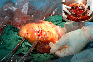
The mass was surgically exteriorized and removed via a midline incision.
The mass was surgically exteriorized and removed via a midline incision.
Image size: 1800 x 1200px
Image size: 1482 x 987px
Fluid was withdrawn from the tumor for hormone assay and to facilitate its removal. Then it was transected as shown in the inset above. The tumor structure was unusual because it consisted of a single large cyst rather that the common multicystic structure of a GCT.
This volume of fluid drained from the tumor was 2200 ml. The fluid was assayed for its estradiol 17 beta, testosterone, progesterone and inhibin concentrations.
Image size: 1800 x 1293px
Serum steroid and inhibin concentrations were (followed by the normal range in each case):
Progesterone: 0.2 ng/ml (0.2 to 10 ng/ml)
Estradiol: 13.8 pg/ml (20 to 45 pg/ml)
Testosterone: 34.4 pg/ml (24 to 45 pg/ml)
Inhibin: 1.56 ng/ml (0.1 to 0.7 ng/ml)
Serum steroid and inhibin concentrations were (followed by the normal range in each case):
Progesterone: 0.2 ng/ml (0.2 to 10 ng/ml)
Estradiol: 13.8 pg/ml (20 to 45 pg/ml)
Testosterone: 34.4 pg/ml (24 to 45 pg/ml)
Inhibin: 1.56 ng/ml (0.1 to 0.7 ng/ml)
Steroid and inhibin concentrations in the cyst were (no normal ranges available):
Progesterone: 0.5 ng/ml
Estradiol: 249.4 pg/ml
Testosterone: 149.0 pg/ml (24 to 45 pg/ml)
Inhibin: 73.8 ng/ml
The
concentrations of serum progesterone, testosterone and estradiol in the serum were consistent with those of a
normal mare. However, the inhibin
concentration in the serum was approximately twice that of a normal mare. These values were consistent with a diagnosis of granulosa cell tumor. This diagnosis was verified by histopathology. The tumor itself was undoubtedly the source of the high concentration of inhibin: the inhibin concentration in the cyst was about a hundred times higher
than its normal concentration in serum.
| In view of the high concentrations of inhibin, it was not possible to explain the presence of the follicles in the contralateral ovary. Also, mounting of a herdmate could not be explained in terms of the pre-operative serum testosterone concentration. |
Note: Low testosterone concentrations are often encountered in mares with GCTs. This underlines its questionable value in the diagnosis of GCTs. It is now (2013) becoming common to measure anti-mullerian hormone (AMH) as a diagnostic aid in these cases.


